May's Case of the Month - 2023
Transitional Cell Carcinoma in a Canine
Patient Information
Age: 7 years
Gender: Spayed Female
Breed: Border Collie Mix
Species: Canine
History
Chronic hematuria. Urinary cultures have been negative. The patient is not responsive to antibiotics.
There is also concern for osteoarthritis or soft tissue injury to the left rear limb, with the patient exhibiting toe-touching to non-ambulatory behavior recently.
Ultrasonographic Findings
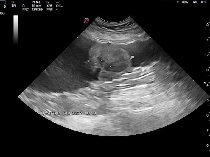
The bladder is moderately distended with anechoic urine and is of relatively normal contour and thickness. There is a 2.8 x 2.3 cm hyperechoic lobular mass originating from the mid-ventral bladder wall. Positive color Doppler noted within the mass.
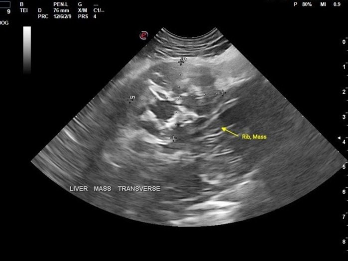
Caudal right rib mass measuring 2.4 x 2.9 cm (sagittal), which appears hypoechoic with multifocal regions of mineralization, located approximately at the 9th–12th rib.
Abdominal Ultrasound Interpretation
Bladder Mass: The findings are moderate to severe.
Differential Diagnoses: Tumors of the urinary bladder are uncommon in dogs and rare in cats. Transitional cell carcinoma (TCC) is the most common primary tumor of the urinary bladder in both species. Other differentials include squamous cell carcinoma, leiomyosarcoma, leiomyoma, rhabdomyosarcoma, or benign bladder polyp. The bladder may also be invaded by prostatic neoplasia or metastatic disease (e.g., hemangiosarcoma, lymphoma). Bacterial cystitis and urolithiasis are common differentials for the clinical signs seen, and a rare but important differential diagnosis for mass lesions is benign polyps of the urinary bladder.
Rib Mass: The findings are moderate.
Differential Diagnoses: Chondrosarcoma, osteosarcoma, soft tissue sarcoma, other neoplasia, less likely granuloma, abscess, or cyst.
Images
Additional Tests
Fine needle aspiration of the suspected rib mass was performed with cytologic interpretation.
Cytology Report
The diagnosis of carcinoma is definitive (100% confidence). Based on the morphology of these epithelial cells with occasional large pink secretory vacuoles, a clinical suspicion for metastatic transitional cell carcinoma is warranted (LeBlanc CJ, Roberts CS, Bauer RW, Ryan KA. Firm rib mass aspirate from a dog. Vet Clin Pathol. 2004;33(4):253-6).
The slides are moderately to highly cellular and consist of a large amount of cellular debris suggesting necrosis, few to many red blood cells, and a nucleated cell population predominated by round to polygonal cells found in cohesive clusters. These cells have a small amount of very basophilic cytoplasm that surrounds a central, round nucleus with a coarsely-stippled chromatin pattern and 1-3 prominent nucleoli. Anisokaryosis is moderate, and binucleation is commonly noted. Occasionally, these cells contain one to two large cytoplasmic vacuoles filled with an abundant amount of thick, pink secretory material. Few leukocytes in proportions consistent with peripheral blood are also present.
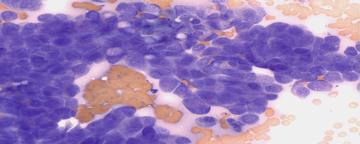
Several clusters of polygonal cells with large round to oval nuclei.
Recommendations
Referral to a veterinary surgeon and/or oncologist for further diagnostics (CT scan) and therapy is highly recommended.
Thoracic radiographs (3-view, metastasis check) are recommended.
Discussion
Invasive transitional cell carcinoma (TCC) is the most common form of canine urinary bladder cancer, affecting tens of thousands of dogs worldwide each year, and its prevalence appears to be rising. Most TCCs are intermediate to high-grade papillary infiltrative tumors; superficial, low-grade tumors are uncommon. TCC is most often located in the trigonal region of the bladder. In one large study of 102 dogs with bladder TCC, more than half of the patients had concurrent urethral involvement, and about a third of male dogs had concurrent prostatic disease. Distant metastases are typically present in about 20% of cases at diagnosis and are associated with a worse prognosis. More than 50% of dogs with TCC have distant metastases by the time of death. Apart from regional lymph nodes and lungs, metastases can occur in the liver, spleen, kidneys, bones, adrenal glands, heart, brain, and skin. The mean age at diagnosis is 9 to 11 years, with females being more commonly affected.
Patient Outcome
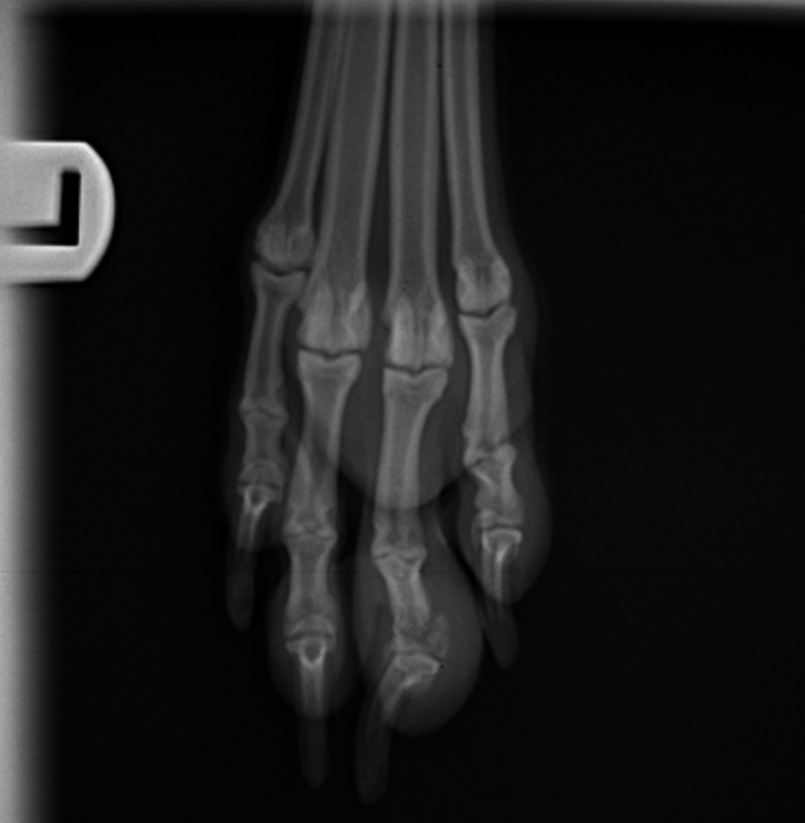
Radiographs of the rear limb showed a left 4th digit distal phalanx fracture with concern for disease infiltration to the bone.
The patient had a scheduled consultation with a veterinary oncologist to discuss therapy options for metastatic transitional cell carcinoma. Approximately one week following the ultrasonographic findings, the patient experienced an episode of collapse and was unable to recover. Emergency care suggested possible metastatic lesions to the spine based on clinical signs. The owner declined further therapy options and elected for euthanasia.
Sonographer: Dr. Mallory Repellin
Special thanks to Wheaton Animal Hospital and Dr. Casey Leblanc at Eastern VetPath.