November's Case of the Month - 2022
Jejuno-Jejunal Intussusception Due to Jejunal Mass
Patient Information:
Age: 8 years
Gender: Spayed Female
Breed: Golden Doodle
Species: Canine
History:
Presented with a two-week history of vomiting and regurgitation. Laboratory work is within normal limits.
Ultrasonographic Findings:
There is a jejuno-jejunal intussusception noted in the left cranial abdomen in the mid-jejunum. Blood flow to entrapped mesenteric fat is adequate. A hypoechoic mucosal vascular mass with punctate hyperechoic shadowing foci is observed at the aboral end of the intussusception, measuring 1.5 x 1.9 cm. The rest of the small intestinal loops exhibit normal bowel layering, thickness, and motility:
- Duodenum: 5.8 mm
- Jejunum: 4.5 mm
- Ileum: 3.9 mm
Minimal luminal dilatation is present orad to the intussusception.
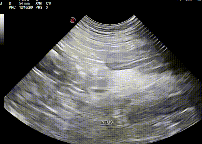
Origin of jejuno-jejunal intussusception.
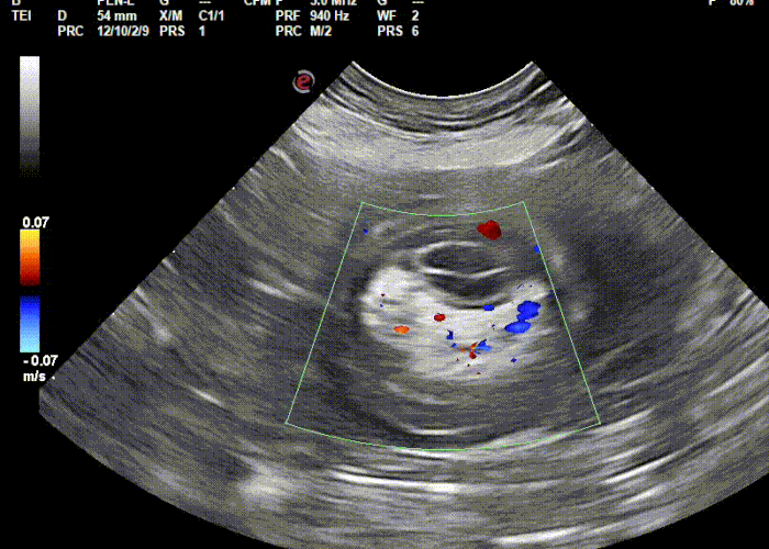
Intussusception in transverse showing normal blood flow to entrapped mesentery.
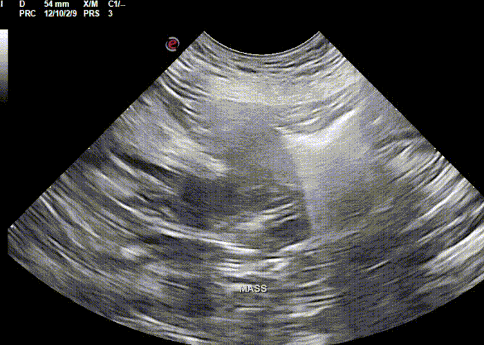
Jejunal Mass present at the termination of the intussusception
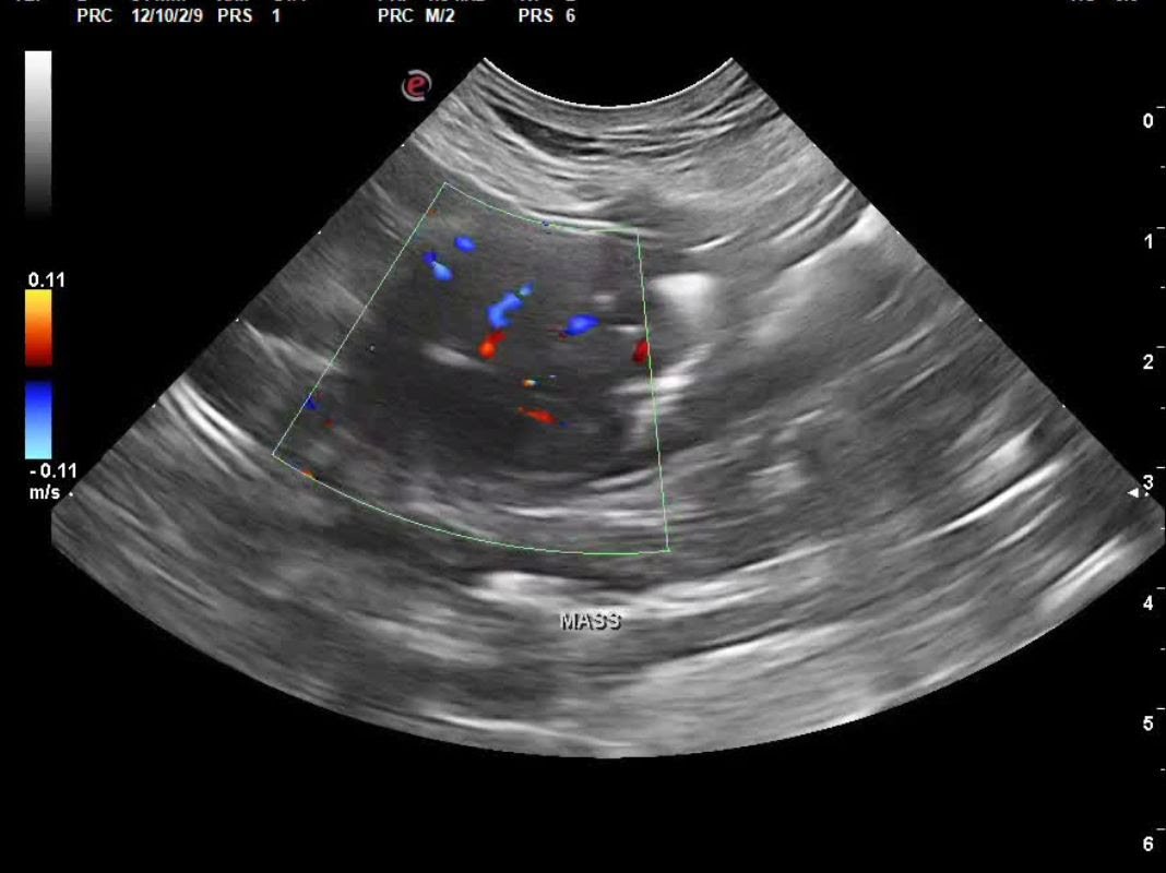
Still image of jejunal mass showing positional color uptake on Doppler exam.
Abdominal Ultrasound Interpretation:
Jejuno-jejunal intussusception - the findings are moderate to severe.
Jejunal Mass - the findings are moderate:
- DDx: adenocarcinoma vs. infiltrative neoplasia (lymphosarcoma vs. mast cell tumor) vs. leiomyosarcoma vs. leiomyoma.
Recommendations:
The prognosis was guarded as the type and behavior of the mass was yet to be determined. A three-view thoracic radiograph and resection with anastomosis, followed by histopathology of the mass, were recommended for an optimal treatment plan.
Outcome:
Unfortunately, due to the guarded prognosis and financial constraints, euthanasia was elected.
Discussion:
Intussusceptions are common in younger animals, with 75% occurring in dogs less than 1 year of age. Any condition causing increased motility can predispose an animal to intussusception. They are often caused by parasitism or enteritis (e.g., parvovirus) but can also result from foreign bodies or intestinal neoplasia. Idiopathic cases are also possible. Clinical signs can be acute or chronic, often including vomiting, diarrhea, and weight loss. The most common type of intussusception is enterocolic, accounting for two-thirds of cases. While radiographs can raise suspicion, ultrasound is the diagnostic modality of choice.
Surgical reduction is usually necessary, with resection and anastomosis required if bowel compromise is present. This procedure often carries a good prognosis if surgical complications do not occur. In this case, hypermotility was likely caused by the presence of a luminal jejunal mass, which caused partial obstruction and accounts for the unusual jejuno-jejunal location of the intussusception.
Sonographer: Kara Woody, DVM
Thank you to Shirlington Animal Hospital for collaborating with us on this interesting case.