
August's Case of the Month 2026 brought to you by Dr. Mallory Repellin.
Case of the month MiVU
Patient information:
Age: 7 months
Gender: Female Intact
Breed: Cane Corso
Species: Canine
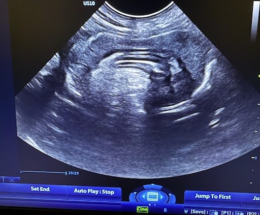
History: Acute onset diarrhea, decreased appetite and lethargy following mass consumption of dirt and suspicion of additional dietary indiscretion. Patient is up to date on DHLPP vaccination and dewormed monthly. She does not associate with unknown dogs, dog parks, or highly social places. Significant Ultrasonographic Findings: Intestinal tract: The stomach is significantly distended with fluid, some gas and dependently-oriented hyperechoic material which does not appear obstructive. Majority of the small intestines are normal-looking loops with mild luminal dilation and no evidence of obstruction to the level of the ileum/ ileocecocolic junction (ICJ). Medial to the right kidney in the normal location of the ICJ there is an abnormal structure with thickened, mildly hypoechoic, homogenous tissue with no appreciated wall layering surrounding subjectively normal loop of small intestine which has adequate wall layering and vascularity. Concerning for intussusception vs. ICJ mass vs. mechanical foreign body (considered less likely)
Mesentery: Mesentery throughout the abdomen, especially in the right mid-abdomen is hyperechoic.
Serosal surfaces: There is a moderate amount of anechoic free-fluid throughout the abdomen.
Image 1: The central intestinal loop with adequate wall layering (intussusceptum) appears contained within a thickened homogenous, well-defined outer segment of bowel (intussuscipiens) to create an irregular image consistent with intussusception but lacks the usual 7-8 layers of intestinal walls generally identified via ultrasonography. The lack of additional wall layers is suspected due to location of the intussusception at the ileocecocolic junction or colon.
Recommendations: Referral of this patient to a veterinary surgeon for emergency surgical intervention is recommended. Concern at this time is for an intestinal obstruction which may be due to intussusception but cannot rule out significant inflammatory, neoplastic, or foreign material-related underlying causes.
Referral to veterinary surgeon (surgery report): - The patient was placed in dorsal recumbency.
Approximately 50-75 ml of clear to slightly straw-colored transudate was noted upon entry into the abdominal cavity. Abdominal exploratory revealed a diffusely and moderately enlarged jejunum with a palpable, firm and long intussusception present at the level of ileocecocolic junction, causing obstruction. The intussusception, centered mostly over the orad portion of the ascending colon, appeared to contain most all of the ileum as well as approximately 7-8 cm of distal jejunum.
An attempt was made to reduce the intussusception, but the intussuscipiens appeared to be completely rolled onto itself, making it nearly impossible to gently milk. Given this unsuccessful attempt, a jejuno-colic resection and anastomosis was routinely performed. The affected segment of small intestine, from 10-12 cm orad to the intussusception (distal jejunum), to 3-4 cm aborad to the intussusception (ascending colon).
Then, a functional end-to-end anastomosis was performed with the remaining healthy bowel. The two ends were apposed side to side (anti mesentery to anti-mesentery) and an endo-GIA stapler was inserted into each intestinal lumen and was locked and fired.
Patient recovered uneventfully from anesthesia.
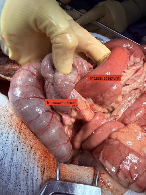
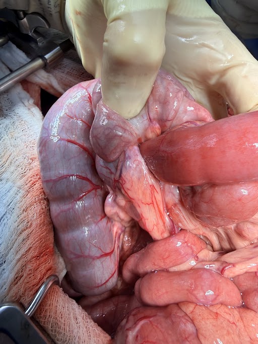
Intraoperative imagery upon identification of the intussusception which is located at the ileocecocolic junction. These images identify both the receiving (intussuscipiens) and telescoped, inner intestinal segment (intussusceptum). The difference in pigmentation depicted in these images is considered normal for both the small and large intestines. Both exposed portions of small and large intestines maintain adequate blood perfusion and coloration at this time.
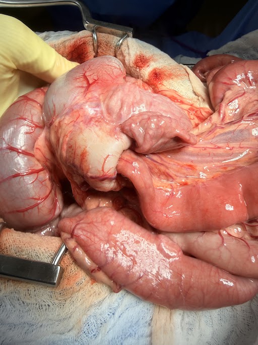
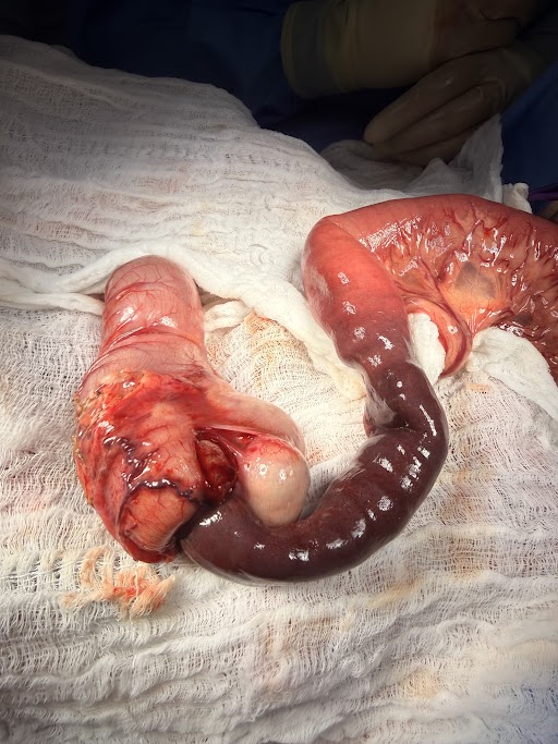
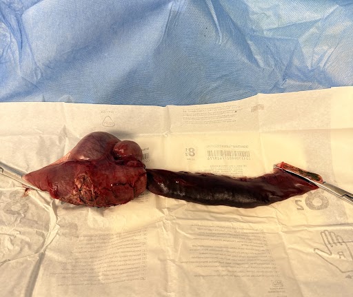
These images shows the site of intussusception where there is blanching of the tissue in the large intestine due to increased internal pressure from the telescoped small intestine. The image on the right shows the purple discoloration of the telescoped inner-portion of small intestine after extraction from within the large intestine. This discoloration is the sequelae of poor perfusion and blood pooling. Intraoperative post-operative images of resection and anastomosis site in situ and following resection
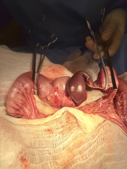
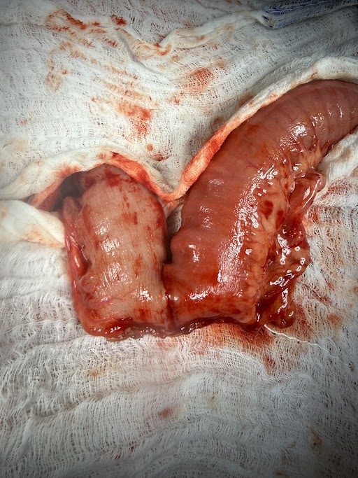
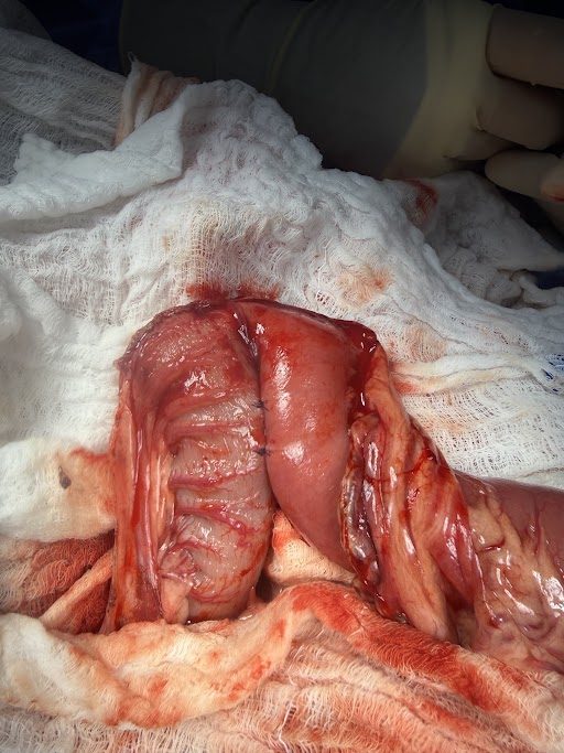
Intraoperative image of post-resected and endo-GIA stapled, re-attached intestines.
Histopathology: Regarding biopsy results being consistent with a completely excised intussusception, with pre-existing mild to moderate lymphoplasmacytic enteritis. There was no evidence of any neoplastic cells or parasites noted. Please refer to attachment for further details. Based on these results, no additional diagnostics or treatments are currently indicated for Bridget, unless clinical signs of diarrhea or vomiting persist (which may be due to enteritis).
Discussion: Intussusception describes a condition in which one segment of the intestine (the intussusceptum) telescopes or invaginates into the lumen of an adjacent segment of intestine (the intussuscipiens).
Intussusceptions may occur at any location in the gastrointestinal tract from the stomach to the large intestine. However, most commonly, the bowel segments involved are the middle of the small intestine (jejunum ) or the where the small intestine joins the large intestine or colon (ileocecocolic junction). Generally the intussusceptum is a more proximal portion of bowel (i.e., closer to the mouth) which telescopes into a more distal (i.e., closer to the anus) segment. This pattern follows the normal direction of peristalsis. The reverse, however, is occasionally found.
Intussusceptions are most commonly associated with some problem that causes inflammation of the intestine (enteritis). Common causes of enteritis are intestinal parasites (hookworms, whipworms, and roundworms), protozoal, bacterial or viral infections (Giardia, Salmonella, canine distemper, and parvovirus) intestinal foreign bodies (bones, plastic toys, etc.), abrupt dietary changes, intestinal masses (tumors), and a surgical procedure performed on the intestine. Increased motility in a segment of intestine (hypermotility) which is adjacent to a segment that has lack of motility (ileus) can cause the hypermotile segment to telescope into the segment with ileus, resulting in an intussusception.
Because most pets that develop intussusceptions have had episodes of vomiting and diarrhea, the hydration and electrolyte status should be addressed prior to surgery if possible. This involves some blood chemistry analysis and treatment with an appropriate intravenous fluid. Treatment of animals with intussusception can be complicated and difficult. Occasionally intussusceptions can be manually reduced by manipulation of the affected bowel through the abdomen or they reduce themselves spontaneously. In most cases, however, surgery is required to treat this problem.
Recurrence of intussusceptions is common, so even if the intussusception can be manually reduced, surgery is often recommended to perform procedures designed to decrease the incidence of recurrence. During surgery the affected area bowel is easily identified. It is occasionally possible for the surgeon to manually reduce the intussusception. In many instances, either the intussusception cannot be reduced or the bowel is so badly damaged that resection of the affected bowel is required. In this case, the area of damaged bowel is removed and the cut ends of the intestine are joined together with sutures or staples in a procedure called an intestinal anastomosis.
Postoperative care following intussusception involves efforts to manage pain, generally with opioids, which help to slow bowel motility as well. Re establishment of hydration and normal electrolyte values is essential and appropriate intravenous fluids are generally used until your pet is eating normally. Antibiotics may be required depending on the amount of contamination from the surgery and the preference of the surgeon.
The prognosis following surgical repair of an intussusception depends on several factors including the duration of the problem, the amount of intestine involved, the location of the problem and the extent of the blockage that has been caused.
Chronic intussusceptions usually require removal of a section of bowel and anastomosis of the ends to re-establish bowel integrity. Anytime bowel has to be removed there is a chance of leakage from the surgery site which can result in potentially fatal peritonitis. Pets that are in poor condition because of the intussusception may have a diminished ability to heal, making leakage more likely. If large amounts of bowel have to be removed, your pet may not do well because of the relatively short length of bowel left behind. The prognosis for pets with an intussusception is good as long as recurrence of the problem can be prevented and excessive amounts of bowel do not have to be removed. It has been reported that between 11% and 20% of dogs will have a recurrence of the problem following surgical correction. The incidence is higher (25%) if only manual reduction and no surgery is done. A procedure known as enteroplication can be performed to prevent recurrence of the intussusception, however, may make the patient more susceptible to other complications such as intestinal obstructions with foreign material that may have been able to pass without complication if the bowel had not been plicated.
Patient outcome: A 2-week post-operative recheck cleared the patient for return to full activity level due to a fully healed ventral abdominal incision.
We are grateful to Fairland Animal Hospital and Bluepearl Veterinary Partners for their cooperation in organizing information on Bridget to tell her story!