
July's Case of the Month 2026 brought to you by Dr. Kara Woody
Feline Infectious Peritonitis Before and After Treatment with GS-441524
Patient Information:
Age: 2 years Gender: Neutered Male
Breed: DSH Species: Feline
History:
Our patient presented with a history of hyporexia and weight loss. Intermittent fever was also noted. Anemia, leukocytosis and hyperglobinemia was present. Possible left renomegaly noted on radiographs.
Abdominal Ultrasonographic Findings 5/5/26:
The right kidney measures 4.0 cm in length with normal shape and normal corticomedullary dimension distinction. No pyelectasia seen.
The left kidney is moderately enlarged measuring 4.8 cm in length with mildly scalloped shape and hyperechoic cortex with normal corticomedullary dimension distinction and a prominent hypoechoic subcapsular "rim." No pyelectasia visualized. Vascularity is normal on Doppler exam.
The spleen is high normal in size (thickness 0.9 cm) with mildly mottled echotexture. No focal lesions appreciated.
The retroperitoneal tissue is severely hyperechoic with hypoechoic striations surrounding the left kidney.
Ultrasound Interpretation 5/5/26:
Left kidney-the findings are severe-DDX: infectious (pyelonephritis) vs toxin (eg. lily toxicosis) vs neoplasia (e.g.lymphoma vs other) vs FIP vs amyloidosis
Spleen (diffuse) - the findings are mild - DDx: neoplasia (mast cell, lymphoma, carcinoma, metastatic disease) vs. fungal (Histoplasmosis) vs. reactive lymphoid hyperplasia vs. splenitis vs. congestion vs. extramedullary hematopoiesis (EMH).
Retroperitoneal tissue - the findings are moderate - DDx: peritonitis - inflammation vs. paraneoplastic reaction vs. infectious vs. fibrosis vs. other.
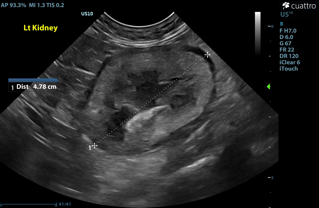
5/5/26-Left Kidney at time of diagnosis
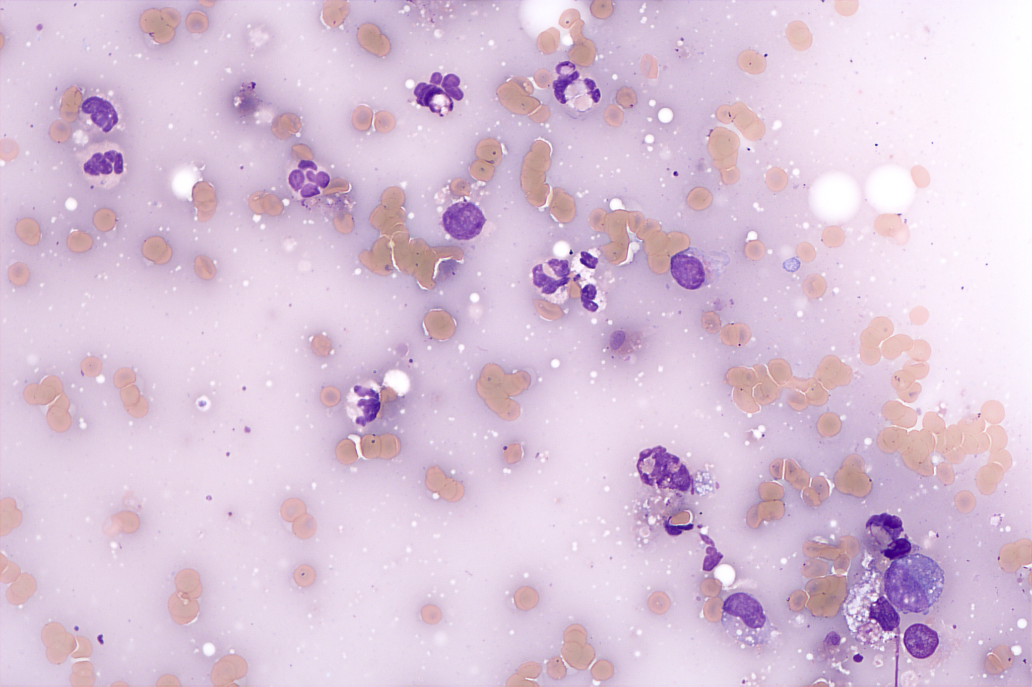
Microscopic image of left kidney cytology showing only low-grade chronic active inflammation; however FIP PCR was positive on these samples. Photo Credit: Eastern Vet Path
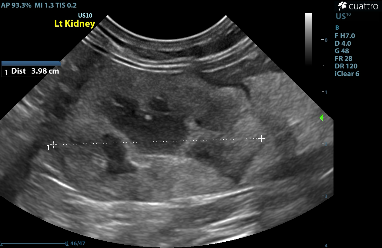
7/6/26-Left Kidney 2 month post treatment. It is now normal size and equal to the right kidney and the hypoechoic sub-capsular “rim” is also noted to be thinner.
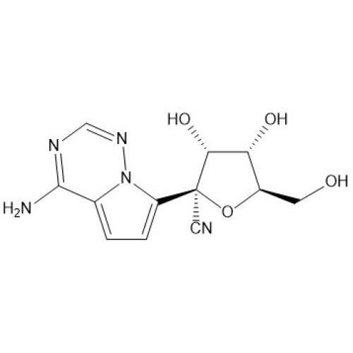
GS-1442454 Chemical structure
Diagnostics:
Sedated ultrasound guided fine needle aspirates of the left kidney were pursued. Cytology findings were suggestive of a low-grade chronic active inflammatory infiltrate, a nonspecific finding with no etiology appearance. FIP PCR was recommended due to high clinical suspicion of FIP with the following results: Auburn Univ Feline Infectious Peritonitis Virus mRNA: POSITIVE (13,200 mRNA Copies per mL Sample) which is consistent with active FIP infection in the sampled organ (left kidney).
Treatment:
GS-441524 was prescribed at 15mg/kg PO daily for 12 weeks. Weight was carefully monitored with appropriate adjustments to account for weight gain. Laboratory work was also checked monthly.
2 month Recheck:
The patient was reportedly doing amazing with normal appetite and weight gain. Laboratory findings had largely normalized with only a mild increase in globulins remaining.
Ultrasound findings 7/6/26:
The kidneys are within normal size limits and symmetrical (Lt/Rt = 4.0/4.0 cm; previously asymmetrical in size 4.8/4.0cm ).
The right kidney has normal size and shape with normal corticomedullary dimension distinction. No pyelectasia visaulized.
The left kidney is smaller (and now normal in size) than previously, but remains mildly scalloped in shape with a thin (previously wider) hypoechoic subcapsular rim. The cortex displays a patchy hyperechoic echogenicity with normal corticomeduallary dimension distinction (unchanged from previous). No pyelectasia visualized.
The spleen is mildly enlarged (thickness up to 1.2 cm; previously 0.9cm) with normal shape and mildly mottled echogenicity. No focal lesions are appreciated.
The retroperitoneal tissue is moderately (previously severely) hyperechoic.
Ultrasound Interpretation 7/6/26:
Left kidney- the findings are moderate and IMPROVED - DDx: resolving changes associated with FIP (likely) vs. chronic inflammatory nephropathy/interstitial nephritis vs. less likely infiltrative disease.
Spleen -the findings are mild and STABLE- DDx: reactive lymphoid hyperplasia vs. splenitis vs. congestion vs. extramedullary hematopoiesis (EMH) vs less likely neoplasia (mast cell, lymphoma, carcinoma, metastatic disease) vs. fungal (Histoplasmosis)
Retroperitoneal tissue - the findings are mild and IMPROVED - DDx: peritonitis - inflammation vs. paraneoplastic reaction vs. infectious vs. fibrosis vs. other.
Patient Outcome:
Given significant improvement in clinical signs, laboratory findings and ultrasound findings, a positive response was made to treatment with GS-441524. Continued improvement is expected.
Discussion:
Feline infectious peritonitis (FIP) is a mutation of the relatively harmless and common feline enteric coronavirus. The mutation allows replication to switch from intestinal cells to monocytes and macrophages, which can then travel through the blood stream and infect other tissues. While FIP is often classified into “wet” and “dry” forms, many infections are actually “mixed.” Clinical signs are usually vague-lethargy, anorexia, fever, diarrhea and sometimes ocular and neurological involvement. Anemia is often present and >50% of cats demonstrate hyperglobinemia. Elevated liver values and azotemia are often noted.
Ultrasound findings can vary in FIP cases. In one study (Muller et al.), effusion was noted in 88% and lymphadenopathy and liver changes in 80%. 68% of cats demonstrated intestinal changes with thickening and layering changes being the most common. Splenic changes (splenomegaly, mottled echotexture) were noted in 36%. Like this patient, renal changes were noted in 32% with most commonly a hypoechoic subcapsular rim and cortical nodules. 92% of cats demonstrated more than one system affected.
Diagnosis can be difficult as feline coronavirus in its non-mutated form is common in cat populations. Fluid analysis of peritoneal effusion can be suggestive of infection, but is not diagnostic. Biopsy and histopathology with immunohistochemical staining for coronavirus antigen in macrophages is the gold standard; however, it can be difficult to perform pre-mortem. The advent of mRNA PCR has made pre-mortem diagnosis easier. The specific test that was used in this patient was performed at Auburn University and detects mRNA of the M gene on the feline corona virus. This demonstrates active replication of the virus in the tissue sampled, which when outside the gastrointestinal tract, is highly suggestive of FIP.
FIP was previously a relatively untreatable, fatal condition. However, in the past few years, development of anti-viral drugs (GS-441524 and its precursors) has changed how we manage these patients with treatment survival rate reported to be 77-96%. GS-441524 competitively inhibits viral replication and is available in oral and injectable forms. Dosage range is typically 10-20mg/kg daily but varies depending on type and severity of infection. Most veterinarians still use the 84 day treatment length. Typically, an improvement in clinical signs is seen in 5-7 days, effusions 1-2 weeks, and globulin levels in 6-10 weeks. Careful and frequent monitoring of clinical signs, body weight, and laboratory findings is recommended during treatment to make sure that dosage adjustments are not needed.
References:
Simons et al. A mRNA PCR for the diagnosis of feline infectious peritonitis. Journal of Virological Methods 124 (2005) 111–116.
Muller et al. Abdominal ultrasonographic findings of cats with feline infectious peritonitis: an update. J Feline Med Surg. December 2023;25(12):1098612X231216000.
Coggins. Updates in Feline Infectious Peritonitis Treatment. ACVIM 2023.
Berliner. Feline Infectious Peritonitis. ACVIM 2019.
Sonographer: Kara Woody, DVM
Special thanks to Toebeans and Tail Veterinary Hospital and Dr. Casey Leblanc with Eastern Vet Path for collaboration on this interesting case with a happy ending!