
June's Case of the Month 2026 brought by Dr. Anne Desrochers
Chronic Hematuria in a Dog
Patient Information
Species: Canine Breed: Labrador Retriever Age: 11 years Sex: Neutered male
History
The patient was presented for evaluation of urinary incontinence and intermittent hematuria of several months' duration. More recently, pollakiuria and stranguria had developed. Digital rectal examination revealed an enlarged prostate.
Ultrasonographic Findings
The prostate was severely enlarged and contained a large, multilobulated, encapsulated intraparenchymal cavitary lesion measuring 2.7×6.6 cm. The cavity contained anechoic to hypoechoic fluid with mobile echogenic particulate material. Multiple hyperechoic foci were visualized adherent to the wall, several producing distal acoustic shadowing, compatible with mineralization and/or gas. The remaining prostatic parenchyma was diffusely heterogeneous and contained a focal hyperechoic area that cast a strong acoustic shadow, consistent with dystrophic mineralization.
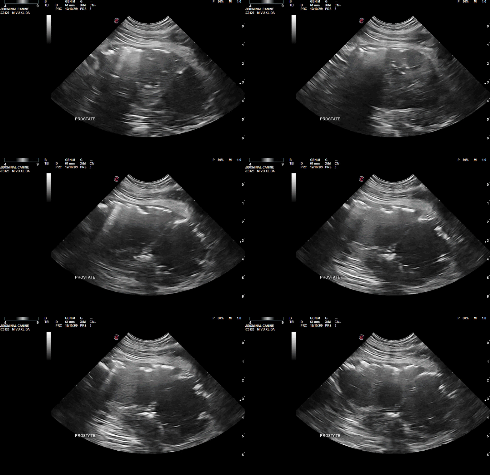
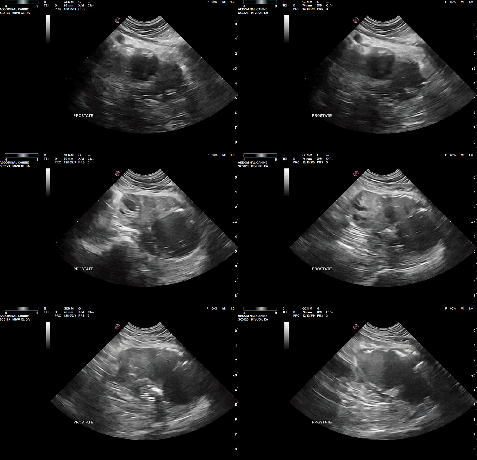
Video 1 & 2. Ultrasonographic cine loops of the prostate demonstrating a large encapsulated cavitary lesion with complex fluid and echogenic intraluminal material, consistent with a chronic prostatic abscess.
Diagnostic Sampling
Ultrasound-guided aspiration of the cavitary lesion was performed for fluid analysis and cytologic evaluation. A fluid sample was also submitted for aerobic and anaerobic bacterial culture and antimicrobial susceptibility testing. Empirical doxycycline therapy was started pending laboratory results.
Cytology revealed abundant necrotic debris, numerous degenerate neutrophils, and extracellular bacterial cocci, consistent with septic suppurative inflammation and abscessation. No cytologic evidence of epithelial neoplasia was identified; however, the possibility of an inflamed or infected neoplasm could not be completely excluded.
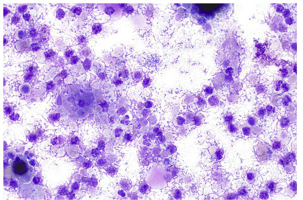
Image 1. Cytology demonstrating abundant necrotic debris, degenerate neutrophils, and bacterial cocci.
Bacterial culture yielded Escherichia coli and β-hemolytic Streptococcus, both susceptible to enrofloxacin and resistant to doxycycline.
Outcome
Antimicrobial therapy was changed to enrofloxacin based on culture and susceptibility results. The owner elected medical management rather than drainage or surgical intervention. Given the patient's neutered status and concurrent intraprostatic mineralization, follow-up ultrasound was recommended to assess treatment response and to rule out an underlying prostatic neoplasm. Due to the recent diagnosis, long-term clinical and ultrasonographic follow-up were not available at this time.
Discussion
Prostatic abscessation represents a severe manifestation of bacterial prostatitis and is most commonly reported in middle-aged to older intact male dogs with concurrent benign prostatic hyperplasia (BPH) or cystic prostatic disease. Ascending urinary tract infection is considered the primary route of bacterial colonization, with Escherichia coli being the most frequently isolated pathogen, although mixed bacterial infections are common. Medical management alone is often associated with treatment failure or recurrence because antimicrobial penetration into large encapsulated abscesses is limited. Consequently, current recommendations favor source control through ultrasound-guided percutaneous drainage or surgical intervention, combined with prolonged culture-directed antimicrobial therapy. Intracapsular prostatic omentalization is generally regarded as the surgical treatment of choice because it provides effective drainage, improves vascular supply, and is associated with lower recurrence rates than historical techniques such as marsupialization. Reported complications include septicemia, abscess rupture, peritonitis, urethral obstruction, and recurrence. Prognosis is generally fair to good when adequate source control and prolonged antimicrobial therapy are achieved but becomes more guarded in cases complicated by systemic sepsis or concurrent prostatic neoplasia. In castrated dogs, prostatic abscessation is uncommon and should prompt consideration of underlying chronic prostatitis, cystic prostatic disease, or prostatic carcinoma, particularly when intraprostatic mineralization is present.
References
1. Johnston SD, Kustritz MVR, Olson PNS. Canine and Feline Theriogenology. Saunders; 2001.
2. Barsanti JA, Finco DR. Disorders of the canine prostate gland. In: Ettinger SJ, Feldman EC, eds. Textbook of Veterinary Internal Medicine. 8th ed.
3. Palmieri C, et al. Canine Prostatic Diseases: An Update. Front Vet Sci. 2022;9:881232.
4. White RAS. Prostatic surgery in the dog. Clin Tech Small Anim Pract. 2000;15:46-51.
5. Smith J. Canine prostatic disease: A review of anatomy, pathology, diagnosis, and treatment. Theriogenology. 2008;70:375-383.
Acknowledgements
Special thanks to the veterinarians and staff at Banfield Pet Hospital–Urbana Pike and Eastern Vet Path for their help with this case.