March's Case of the Month - 2026
Urinary Neoplasia and Perinephric Pseudocyst in a Cat
Patient Information
Age: 18 years
Gender: Neutered Male
Breed: Maine Coon Mix
Weight: 10.5lbs
History
Patient was presented for ultrasound due to weight loss, hyporexia, and an abdominal mass palpated during physical exam.
Abnormal abdominal ultrasound findings -
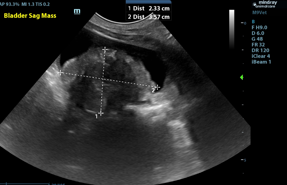
Bladder:
- The bladder is moderately distended with anechoic urine with a 2.3 x 3.6cm hyperechoic heterogenous mildly mineralized mass in the bladder that is sitting in the region of the trigone and occupying a moderate amount of space in the bladder lumen.
Kidneys and Ureters:
- Both kidneys are mildly enlarged (Lt/Rt = 4.7/4.7cm ) and normal shape with coarse moderately hyperechoic renal cortices which are disproportionately large. There is moderate loss of the corticomedullary junction distinction. There is a severe amount of renal pelvic dilation in both kidneys (Lt/Rt - 8.7/8.7mm) with a moderate amount of dilation in the diverticula.
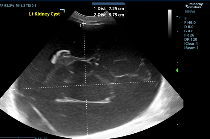
- There is a severely enlarged cystic structure (at least 7.3x9.8cm) surrounding the left kidney that is filled with mildly echogenic fluid with thin, hyperechoic strands extending through the cavitary region creating a septate appearance. Some of these strands connect to a 2.0x0.6cm hyperechoic heterogenous collection of tissue that sits along the capsule of the left kidney.
- There is a mild amount of anechoic fluid around the right kidney.
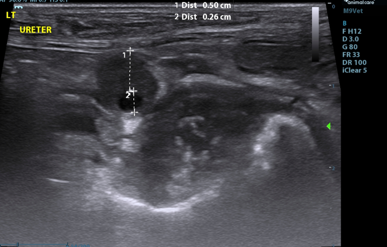
- Both ureters are mildly to moderately dilated along their length (Left - up to 4.3mm (decreased to 2.5mm at the level of the trigone); (Right up to 3.1mm - (decreased to 1.9mm at the level of the trigone)).
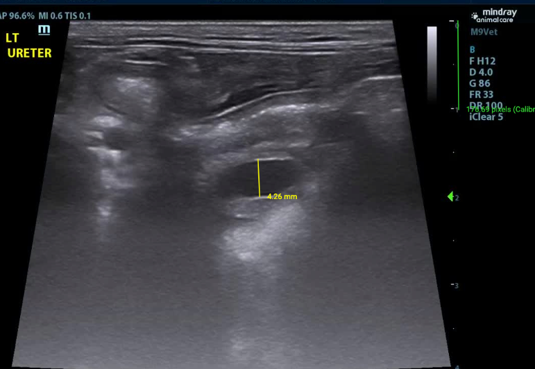
- The walls of the left ureter are mildly to severely and disproportionately thickened along its length (up to 5.0mm)
Serosal Surfaces:
- A mild amount of anechoic free fluid is seen throughout the abdomen.
Differentials
Bladder Mass - the findings are moderate - DDx: Tumors of the urinary bladder are uncommon in dogs and rare in cats. Transitional cell carcinoma (TCC) is the most common primary tumor of the urinary bladder in both species. Other differentials include squamous cell carcinoma, leiomyosarcoma, leiomyoma, and rhabdomyosarcoma or benign bladder polyp. The bladder may also be invaded by prostatic neoplasia or metastatic disease (e.g., hemangiosarcoma, lymphoma).
Ureteral thickening - the findings are moderate - Ddx: ureteral wall neoplasia (ascended from bladder neoplasia) vs. ureteritis
Kidneys (diffuse changes) - the findings are moderate - DDX:
a) Chronic nonspecific change - (chronic glomerulonephritis vs. amyloidosis), chronic interstitial nephritis, chronic nephritis. In cats, the loss of corticomedullary distinction is not unusual with chronic renal disease as interstitial fibrosis in the medulla renders its echogenicity similar to that of the cortex.
b) Acute renal failure/Nephritis (infectious, GN, toxic, etc.) vs. Acute-on-Chronic renal failure
c) Lymphosarcoma
d) Pyelonephritis
Hydronephrosis - the findings are moderate - DDx: Pyelonephritis and ureteritis vs. Increased diuresis caused by renal insufficiency or other condition vs. Toxin vs. Infectious (leptospira, etc) vs. Post-renal obstruction / Bladder distention
Cystic Structure around Left Kidney - the findings are severe and consistent with a perirenal/perinephric pseudocyst, most commonly associated with chronic interstitial kidney disease, neoplasia, prior trauma, and urethral obstruction. Other differentials include renal neoplasia or renal abscess
Ascites- this finding is mild - DDx: transudate vs. hemorrhagic vs. exudate
Recommendations and Outcome
- Referral for oncology and/or surgery consultation was recommended, however due to the severity of the sonographic findings, palliative care was recommended if further diagnostics and treatment were not pursued. Due to the patient’s poor prognosis, the owners elected humane euthanasia.
Discussion
- Given the sonographic findings, partial obstruction of the ureters by the trigone mass with invasion of primary bladder neoplasia up the left ureter wall was most strongly suspected. The perinephric pseudocyst could have existed prior to bladder disease or could have developed secondarily which is considered more likely.
- A perinephric pseudocyst is a collection of a large volume of fluid around a kidney.
- True cysts by definition are lined by epithelium, which is not the case with these collections of fluid which are typically subcapsular (though some extracapsular have been reported), hence the name pseudocyst.
- There is no understood cause of perinephric pseudocysts, however they have been associated with several underlying renal diseases including neoplasia, hydronephrosis, interstitial nephritis, and polycystic renal disease.
- Treatment for perinephric pseudocysts is surgical capsulectomy/fenestration (+/- omentalization)
Citations
Mattoon, John S., Rance K. Sellon, and Clifford R. Berry, editors. Small Animal Diagnostic Ultrasound. 4th ed., St. Louis, MO: Elsevier, 2021.
Special thanks to Stonehenge Veterinary Hospital for their collaboration on this interesting case