
October's 2026 Case of the Month brought to you by Dr. Rebecca Barnes!
“Sugar High”: Uncontrolled Diabetes Mellitus in a Senior Cat
Patient Information:
Age: 7 years
Gender: Neutered Male
Breed: DSH
HISTORY:
April 2026: Diagnosed with Diabetes Mellitus . Over the last few months, he has experienced a rapid weight gain of at least 4 lbs. There has been no change in his diet, which consists of Iams regular adult and Friskies. He has an increased appetite and cries frequently as if seeking food.
July 2026: Presented for vomiting of 24 hours duration. BG 424. Insulin 2U BID. Diagnosis: acute vomiting with uncontrolled diabetes mellitus
August 2026: Increased insulin to 3U BID, then 4U in the morning, 3U in the evening based on blood glucose monitoring at home (BG 364).
September 2026: Presented for diarrhea of 5 days duration. Insulin remains at 4U in the morning, 3U in the evening. BG 335. Diagnosis: Pancreatitis. Abdominal ultrasound recommended to further evaluate the underlying cause of pancreatitis and assess for causes of difficulty controlling the patient’s diabetes.
PHYSICAL EXAM FINDINGS:
Unremarkable physical exam findings, apart from increased BCS secondary to weight gain
LABORATORY ABNORMALITIES:
4/2026: Initial diagnosis of Diabetes Mellitus
GLU 225 mg/dL H (72.00 - 175.00)
7/2026: Acute vomiting episode
GLU 424 mg/dL H (72.00-175.00)
8/2026: Routine diabetes recheck
GLU 364 mg/dL H (72.00-175.00)
9/2026: Diagnosis of pancreatitis
Pancreatic Lipase >50.0 H (0.0-4.4)
ULTRASOUND FINDINGS
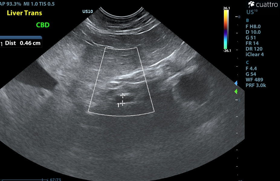
The liver is moderately enlarged with rounded margins and diffuse hyperechogenicity. No focal lesions are appreciated. The gallbladder is moderately distended with normal anechoic and hyperechoic unorganized dependent bile that is not resulting in obstruction. Mild common bile duct dilation is present, measuring 4.6 mm (normal < 4.0 mm) (Image 1).
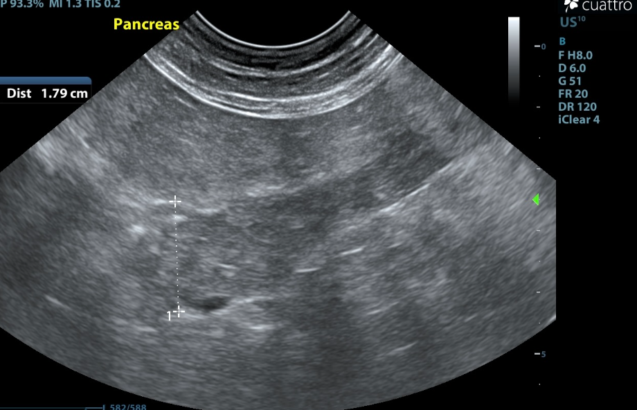
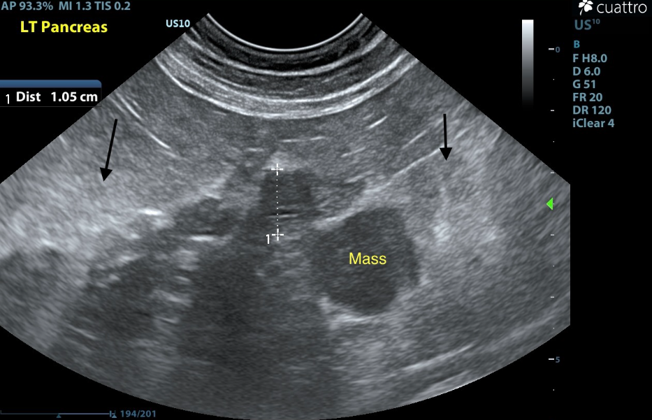
The pancreas is enlarged, hypoechoic, irregularly marginated, and coarse, measuring up to 1.8 cm thickness. Peripancreatic fat is hyperechoic. No focal lesions seen (Image 2).
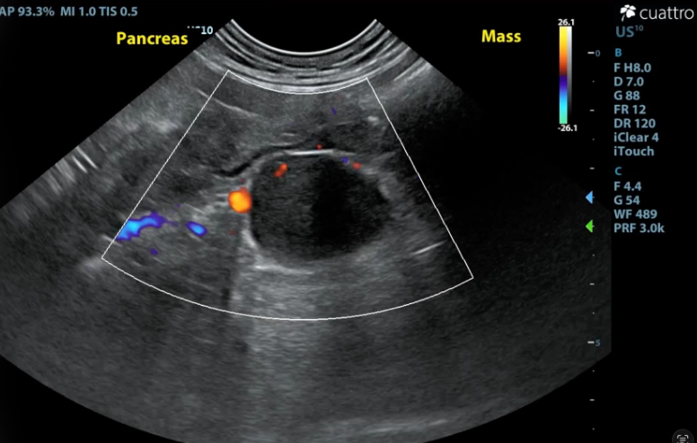
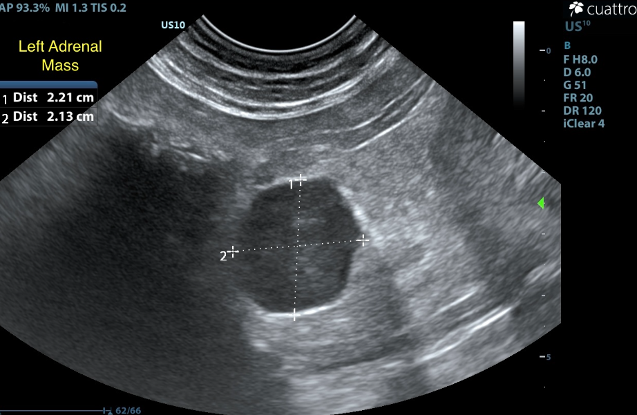
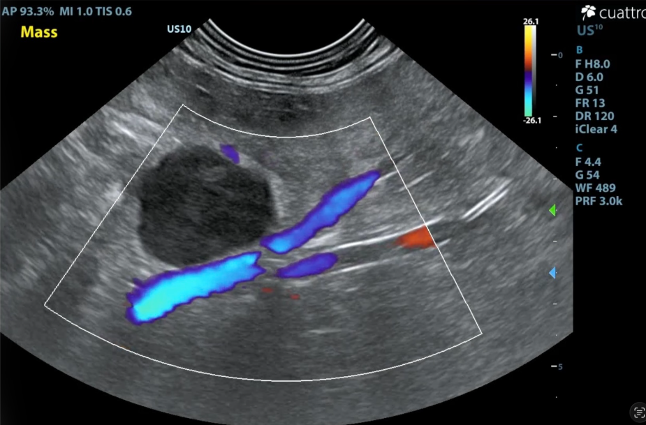
The right adrenal gland was visualized and recognized as having normal shape, size (Rtcd - 3.7 mm), position, and echogenicity. A homogeneously hypoechoic left adrenal mass is present abutting the left pancreatic limb measuring 2.2 x 2.1 cm (Image 3 and 4). There is mild mesenteric hyperechogenicity surrounding the mass which may represent the peripancreatic fat v. focal steatitis around the mass itself (Image 5). No adrenal invasion in the vena cava, phrenic vein thrombosis, dystrophic mineralization or clinically significant nodular changes were noted (Image 6).
ULTRASOUND INTERPRETATION
Liver (diffuse): moderately enlarged and hyperechoic - DDx: Steroid hepatopathy / Vacuolar hepatopathy, Diabetes Mellitus, Chronic vs. Acute hepatitis or cholangiohepatitis (bacterial vs. sterile vs. toxin), less likely infiltrative disease (lymphoma).
Common bile duct: mild dilation - DDx: secondary/reactive change associated with pancreatitis, partial extrahepatic biliary obstruction, cholangitis
Pancreas: moderate pancreatic enlargement/coarse echotexture with hyperechoic peripancreatic fat - DDx: pancreatitis (acute, chronic, acute-on-chronic), pancreatic neoplasia, less likely peripancreatic fat necrosis
Left adrenal gland: left adrenal mass - DDx: Cortical mass-adenoma vs adenocarcinoma (functional vs non-functional). Feline functional cortical masses can secrete more than one hormone (aldosterone, cortisol, and/or sex hormones). Medullary mass-pheochromocytoma-rare
DISCUSSION
Adrenal masses in cats are rare, and make up about 0.2% of all feline tumors. Masses are categorized as cortical or medullary. Cortical masses arise from the outer layer of the adrenal gland and include benign adenomas or malignant carcinomas. They can be functional (producing excess hormones) or non-functional. Medullary masses (Pheochromocytomas) arise from the inner core and secrete stress hormones like catecholamines.
Functional adrenocortical masses produce aldosterone, cortisol, and/or sex hormones (androgens, progesterone, estrogen) in excess. Clinical signs and laboratory findings vary depending on which hormone, or hormones, are secreted and have a fair amount of overlap. Aldosterone secretion (primary hyperaldosteronism), can cause hypokalemia which would lead to muscle weakness, plantigrade stance, and/or neck ventroflexion, as well as high blood pressure, and potential vision loss or retinal detachment. Excess cortisol (hyperadrenocorticism) can cause diabetes mellitus, lethargy, muscle weakness, and thin skin. Sex hormones (androgens, progesterone, estrogens) can cause behavioral changes such as inappropriate urination/spraying, strong-smelling urine, inter-cat aggression, excessive vocalization, diabetes mellitus, or fragile skin syndrome.
Medullary masses (Pheochromocytomas) are rare, and cause excess production of catecholamines. Clinically these cats can have rapid heart rate, high blood pressure, and potential cardiovascular distress.
An adrenal mass may be suspected based on these physical exam findings and lab work abnormalities. But, would likely fall lower on an initial differentials list as most of these findings are overlapping and nonspecific, and can be indicative of many medical conditions. Imaging (ultrasound or CT), when deemed clinically appropriate, is needed to confirm the presence of an adrenal mass. Adrenal masses are often not readily identified on radiographs. Once a mass is confirmed on imaging, a low-dose dexamethasone suppression test or urine cortisol-to-creatinine ratio, help evaluate for hyperadrenocorticism (HAC) (cortisol-secreting mass) and additional adrenal steroid profiles may be used to detect excess aldosterone, progesterone, and/or other hormones.
Link Between Adrenal Masses and Diabetes
Cortisol and/or progesterone-secreting adrenocortical masses cause severe insulin resistance, leading to secondary diabetes mellitus in about 80% of affected cats.
In cases of adrenal-dependent hyperadrenocorticism (cortisol-secreting adrenocortical mass), excess cortisol acts as an anti-insulin hormone. It decreases cellular glucose uptake and ramps up glucose production in the liver. Therefore, cats with adrenal-dependent hyperadrenocorticism, and secondary diabetes mellitus, often require very high doses of insulin, or their diabetes remains completely uncontrolled despite standard treatment.
Progesterone, similar to cortisol, acts as an antagonist to insulin, blocking its normal ability to move glucose out of the blood and into cells. Cats with a progesterone-secreting adrenocortical mass, and secondary diabetes mellitus, require large and/or rapidly increasing doses of insulin without achieving glucose control.
Treatment and Outlook
Unilateral adrenalectomy is the treatment of choice for functional adrenal masses. This removes the source of excess hormone secretion, and allows for tissue sample submission for histopathologic evaluation.
If surgical excision is not an option, medical management would be aimed to control the underlying excess hormone secretion. For aldosterone-secreting masses, medical management includes spironolactone to block aldosterone, potassium supplementation to combat hypokalemia, and antihypertensive medications to control blood pressure. Cortisol-secreting masses (hyperadrenocorticism), are treated with Trilostane.
For patients with secondary diabetes mellitus, once the underlying adrenal mass is successfully removed or medically managed, insulin resistance often drops, and some cats enter complete diabetic remission within several months
OUTCOME
10/2026:
Potassium = 2.9 mmol/L L (3.5 - 5.8)
GLU = 287 mg/dL H (74 - 159)
LIPA = 1279 U/L (100 - 1400)
Blood pressure results: 181/109 map 152, 188/109 map 133, 175/122 map 139
A follow-up chemistry panel with electrolytes was performed after the adrenal mass was found on ultrasound. This panel showed continued hyperglycemia, despite increases to the insulin dose, with a normalized lipase. Additionally, hypokalemia was noted on the electrolyte panel. These findings raised concern that this patient’s adrenal mass is indeed functional and may be secreting multiple hormones, such as aldosterone along with cortisol and/or progesterone, given his secondary, difficult-to-control diabetes mellitus, hypokalemia, and mild hypertension.
Additional endocrine testing and/or plans for surgery have not yet been pursued at the time of this report.
References
Daniel et al. Journal of Feline Medicine and Surgery (2015) 77-84. Clinical findings, diagnostics and outcomes in 33 cats with adrenal neoplasia (2002-2013). https://pmc.ncbi.nlm.nih.gov/articles/PMC11149020/
Cook and Evans. Journal of Feline Medicine and Surgery (2021) 4-16.
Feline comorbidities: Recognition, diagnosis, and management of the cushingoid diabetic. https://pmc.ncbi.nlm.nih.gov/articles/PMC11163885/
Williams. VCA Animal Hospitals. Cushing’s Disease in Cats. https://vcahospitals.com/know-your-pet/cushings-disease-in-cats
UTCVM Veterinary Medical Center, Diagnostic Laboratory Service, Endocrinology. https://vetmed.tennessee.edu/vmc/dls/endocrinology/
Thank you to Seaside Animal Hospital for collaborating with us on this case.