
September's Case of the Month 2026 brought to you by Dr. Emily Evans
An unexpectedly hairy situation
SIGNALMENT:
Breed: DSH
Sex: Neutered Male
Age: 3.5y Weight: 12.3lbs
HISTORY:
Intermittent emesis was reported among the household cats over recent weeks, with the specific individual previously unidentified. This patient was subsequently observed vomiting and exhibiting diarrhea. The evening preceding ultrasonographic examination, the patient experienced six episodes of emesis and displayed marked hyporexia, representing a distinct acute decline in an individual with a historically excellent appetite.
Serum biochemistry and hematology revealed mild hemoconcentration consistent with dehydration.
ULTRASOUND FINDINGS:
The stomach had normal wall thickness and layering. The stomach contained a mild amount of non-obstructive digest and a mild amount of gas. The pylorus was free of obstruction. A mild amount of gas was noted in the small intestine. Small intestinal loops had normal to mildly increased wall thickness with preserved layering. One jejunal loop contained a non-obstructive shadowing intraluminal structure.(4.5x1.7mm) No excessive dilation, plication, obstructive foreign material, or masses were seen. The colon had normal wall thickness and layering throughout. Duodenum-4.0 mm; Jejunum-1.7-2.7 mm; Ileum 3.8mm, Colon-1.2 mm
The mesenteric lymph nodes were mildly enlarged and hypoechoic(up to 6.5mm).
No free fluid, focal lesions or changes in mesenteric echogenicity noted. There was a hyperechoic rounded shadowing nodule in the cranial right abdomen. (0.6cm)
ULTRASOUND IMPRESSIONS:
Intestines - mild - DDX: infectious enteritis vs. secondary dietary indiscretion vs. inflammatory bowel disease/food intolerance vs. infiltrative neoplasia (small-cell lymphosarcoma vs. mast cell tumor) vs. parasitism (cestode) vs. dry FIP vs. fungal (histoplasmosis). Inflammatory bowel disease in cats can be chronic and long-standing but in many cases will transition into small-cell lymphosarcoma and it is not possible with ultrasound alone to determine in this case whether the disease is benign or infiltrative.
Shadowing non-obstructive jejunal luminal structure - mild -DDx: transient foreign material vs. normal dense digest.
Mesenteric lymph nodes: mild enlargement and hypoechogenicity, likely reactive - DDx: reactive lymphadenopathy secondary to gastrointestinal inflammation, infectious/inflammatory enteropathy, FIP, infiltrative neoplasia
Cranial right shadowing structure - mild - incidental findings- DDx: bates body(suspected) vs. other dystrophic mineralization.
PLAN -
Continue conservative GI therapy and consider recheck ultrasound in 4 days while MIVU was back in the clinic. There was minimal improvement over the next 4 days.
RECHECK ULTRASOUND (4 Days after initial ultrasound- GI Focused)
The stomach contained a moderate amount of acoustic shadowing ingesta. (~4cm in length)The visible gastric walls had normal thickness and layering. The pylorus was obscured by shadowing material in the stomach. The proximal duodenum was mildly thickened with no plication or shadowing luminal material.
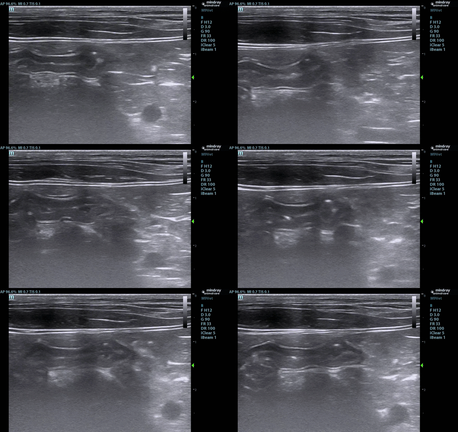
Some loops of jejunum had asymmetric thickening with mucosal layer thickening. In the right cranial quadrant there was a loop of small intestine with marked corrugation/mild plication. Marked motility was noted in the loop and a single thin hyperechoic linear structure is noted passing through the affected intestine. This area was dynamic in appearance and would relax to a less plicated appearance. There was a loop of small intestine that had an acoustic shadowing poorly defined non-obstructive structure in the lumen. (2.3mm in length) Small intestine measurements 3.1-3.6mm
The colon had normal wall thickening and layering.
Mild mesenteric and ICCJ lymph nodes were hypoechoic and plump.
A trace amount of free fluid was noted. The mesentery was mildly hyperechoic in echogenicity with no focal areas of change.
There is a hyperechoic rounded shadowing nodule in the cranial right abdomen. (0.6cm)
RECHECK ULTRASOUND IMPRESSIONS:
Stomach/Intestines - the findings were moderate and progressive and suggestive of an intraluminal gastric foreign body, possible non-obstructive intestinal foreign material, and developing linear foreign body.
Intestine(thickening and layering change) - the findings were mild-moderate and progressive - DDX: Inflammation secondary to passing foreign material/developing obstruction vs. inflammatory bowel disease/food intolerance vs. inflammation secondary to foreign material vs. infiltrative neoplasia (small-cell lymphosarcoma vs. mast cell tumor) vs. parasitism (cestode) vs. dry FIP vs. fungal (histoplasmosis)
Lymph nodes - the findings were mild and stable - DDx: reactive likely vs. infection vs. IBD vs. unlikely - infiltrative neoplasia (lymphoma vs. mast cell vs. other) vs. metastatic neoplasia
Ascites- this finding were trace and new - DDx: transudate vs. hemorrhagic vs. exudate
Bates Body- Stable incidental findings
Image 1- Hyperechoic Thin Linear Structure in loop of bunching small intestine
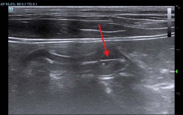
Image 2: Hyperechoic Thin Material in Small Intestine Loop
EXPLORATORY LAPAROTOMY AND FINDINGS:
Given the progression of sonographic findings and concerns for linear foreign material coupled with minimal improvement clinically. Exploratory laparotomy was performed. A moderate sized
trichobezoar was found in the stomach and was removed. A smaller Trichobezoar was attached to a single strand of human hair causing plication was located in the jejunum.
Image 3: Strand of hair found causing intestinal plication
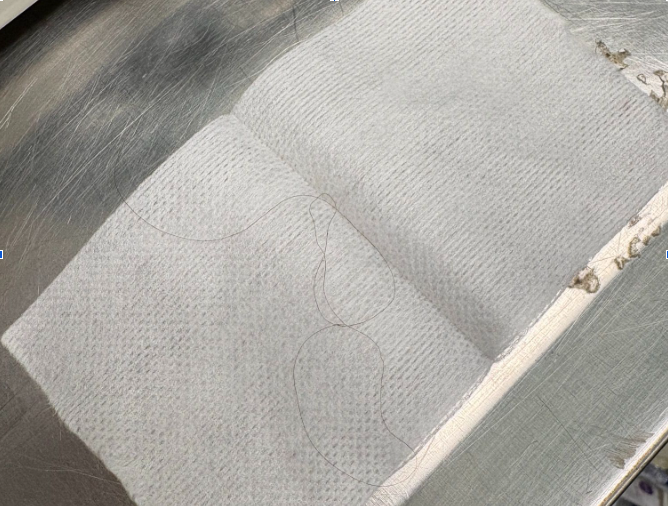
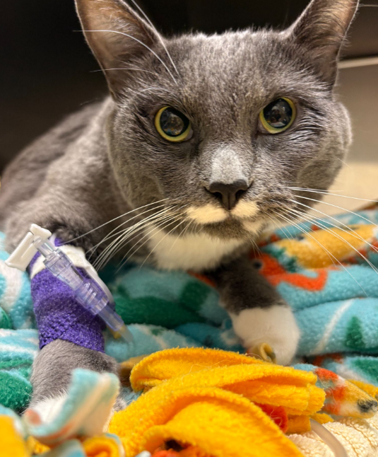
Image 4: Patient recovering comfortably post operatively
DISCUSSION:
Discrete intraluminal foreign bodies are typically distinct and sonographically evident, often producing acoustic shadowing alongside proximal intestinal distension, hyperperistalsis, and complete post-obstructive bowel collapse. In contrast, linear foreign objects frequently present as a thin, hyperechoic intraluminal structure traversing plicated, corrugated loops of small intestine. Distinct acoustic shadowing is generally absent with fine materials such as thread or hair, whereas prominent shadowing typically occurs with thicker fabrics.
Initial ultrasonographic evaluation of the patient’s gastrointestinal tract revealed no overt obstruction or secondary changes characteristic of linear foreign material that would necessitate surgical intervention. However, sequential ultrasonography demonstrated disease progression, enabling improved visualization of a gastric trichobezoar and an associated strand of hair inducing early intestinal plication. A recent retrospective study investigating feline gastrointestinal foreign bodies indicates that the overall prognosis for cats undergoing exploratory laparotomy for discrete or linear foreign bodies is favorable, with a low incidence of postoperative complications or mortality.
The patient has made an uncomplicated recovery following surgery, rapidly regaining his excellent appetite.
REFERENCES:
[1] Barrs VR, Beatty JA, Tisdall PL, Hunt GB, Gunew M, Nicoll RG, Malik R. Intestinal obstruction by trichobezoars in five cats. J Feline Med Surg. 1999 Dec;1(4):199-207. doi: 10.1053/jfms.1999.0042. PMID: 11714236; PMCID: PMC10822377.
[2] Cannon M. Hair balls in cats: a normal nuisance or a sign that something is wrong? J Feline Med Surg. 2013 Jan;15(1):21-9. doi: 10.1177/1098612X12470342. PMID: 23254238; PMCID: PMC10816490.
[3] Gollnick HR, Schmiedt CW, Wallace ML, Sutherland BJ, Grimes JA. Retrospective evaluation of surgical treatment of linear and discrete gastrointestinal foreign bodies in cats: 2009-2021. J Feline Med Surg. 2023 Jun;25(6):1098612X231178140. doi: 10.1177/1098612X231178140. PMID: 37350262; PMCID: PMC10811983.
[4] Lindquist E, Casey D, Frank J. Sonographic criteria for the diagnosis of gastrointestinal obstruction in 39 dogs and cats. SonoPath.com and Sound Technologies New Jersey Mobile Associates, Sparta, NJ, USA.
Thank you to Ashland Animal Hospital for trusting us to help one of your own!