Severe congenital kidney disease in a young cat
Patient information:
Age: 1 year, 9 months
Gender: Male Neutered
Breed: DSH
Species: Feline
History:
Per the referring veterinarian, this patient was found to have elevated kidney values which are steadily and progressively increasing despite fluid therapy. An abdominal ultrasound was recommended for further investigation.
Significant Ultrasonographic Findings:
Liver:
Normal size, shape and patchy mixed echogenicity with coarse echotexture. The hepatic vasculature appears dilated with additional, possibly anomalous branches appreciated. No focal lesions are appreciated. The gall bladder is moderately distended with normal anechoic and hyperechoic unorganized dependent bile that is not resulting in obstruction. No common bile duct dilation is seen.
Kidneys:
Severely enlarged size (Lt/Rt = 6.9/6.0 cm) and normal overall shape with obliteration of normal renal architecture, there are TNTC variably sized anechoic cystic lesions throughout both kidneys. Adequate vasculature to the kidneys is appreciated. Bilateral pyelectasia visualized (Lt/Rt= 0.4/0.3 cm).
Serosal surfaces:
Mesentery throughout the abdomen is diffusely hyperechoic.
Abdominal ultrasound interpretation:
Renomegaly - the findings are severe - DDX: polycystic kidney disease, unlikey infectious (pyelonephritis) vs toxin (eg. lily toxicosis) vs neoplasia (e.g.lymphoma vs other) vs FIP vs amyloidosis
Liver - the findings are mild - DDx:
Steroid hepatopathy / Vacuolar hepatopathy / Glycogen storage disease
Chronic vs. Acute hepatitis or cholangiohepatitis (bacterial vs. sterile vs. toxin)Diabetes
mellitus
Infiltrative neoplasia (lymphosarcoma)
Fungal infection
Hepatotoxin
Mesentery - the findings are mild - DDx: peritonitis - inflammation vs. paraneoplastic reaction vs. infectious vs. fibrosis vs. other.
Images:
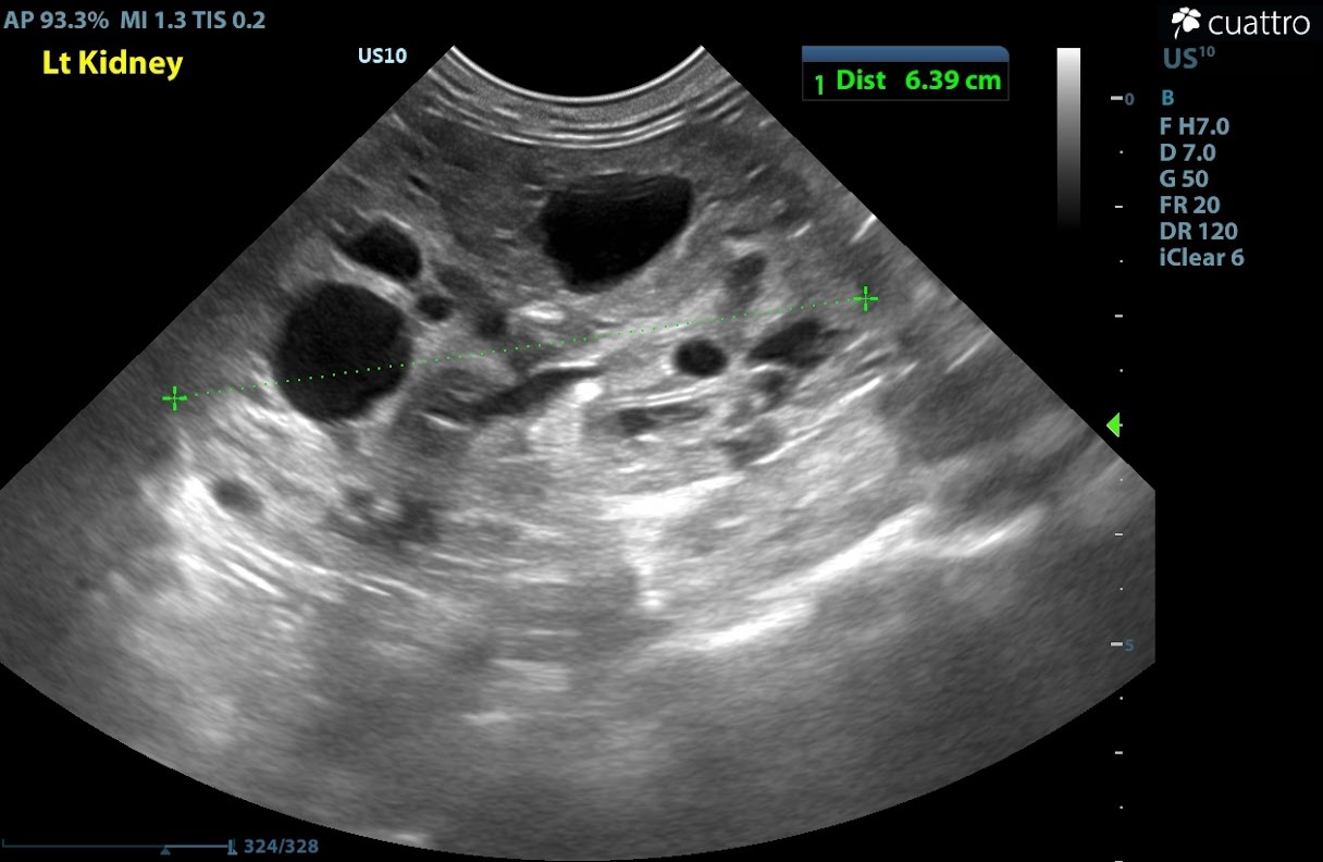
Sagittal view of the left kidney. Normal feline renal length is 3.2-4.2 cm. The left kidney appears to have enlarged size and overall normal shape, a significant number of anechoic cortical cystic lesions are present throughout the kidney.
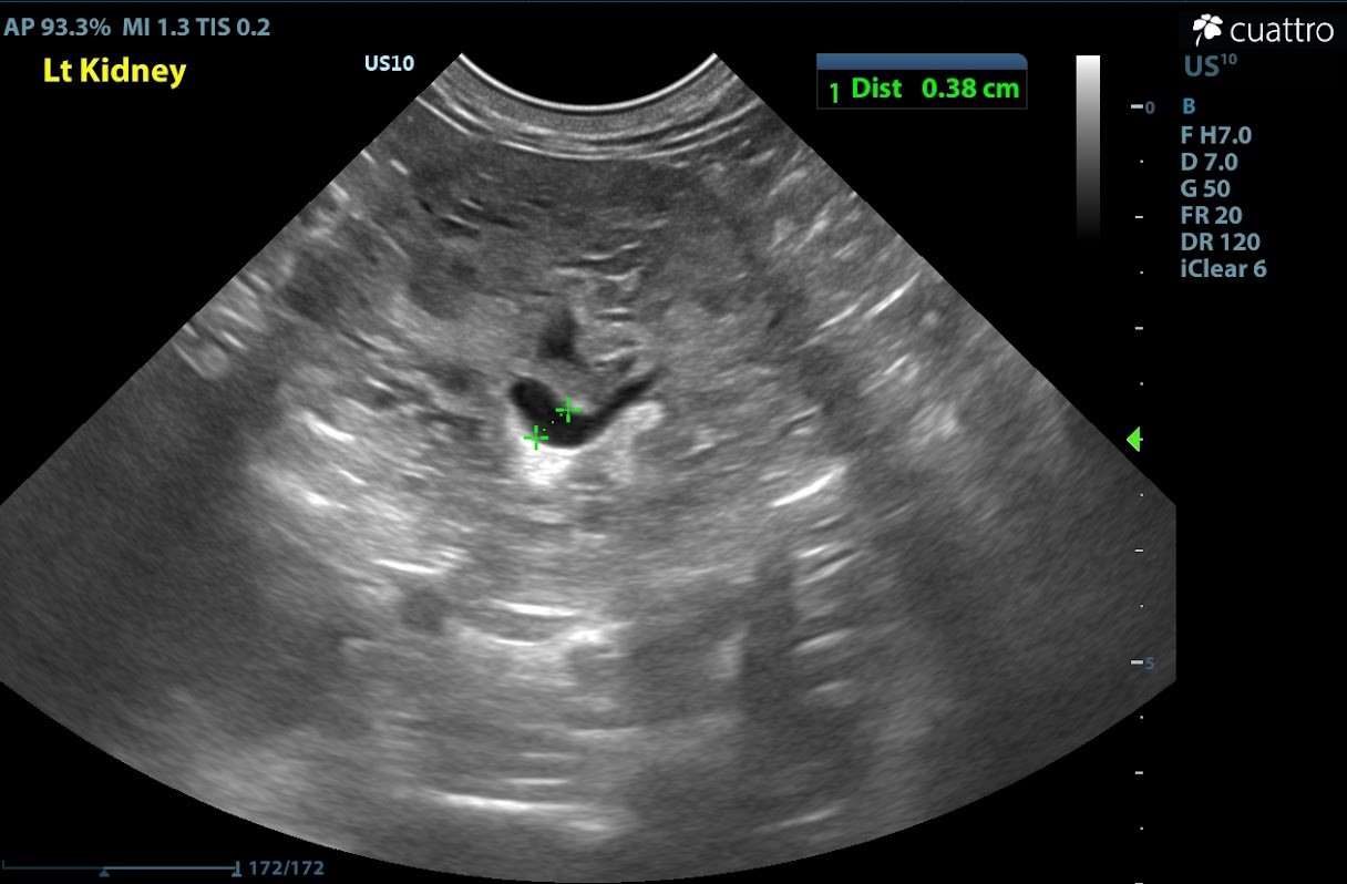
Transverse orientation of the left renal pelvis demonstrating dilation (i.e. pyelectasia) which indicative of a direct insult to the kidney. This finding can be appreciated in acute and chronic renal disease, ascending urinary tract infections, and ureteral obstructions.
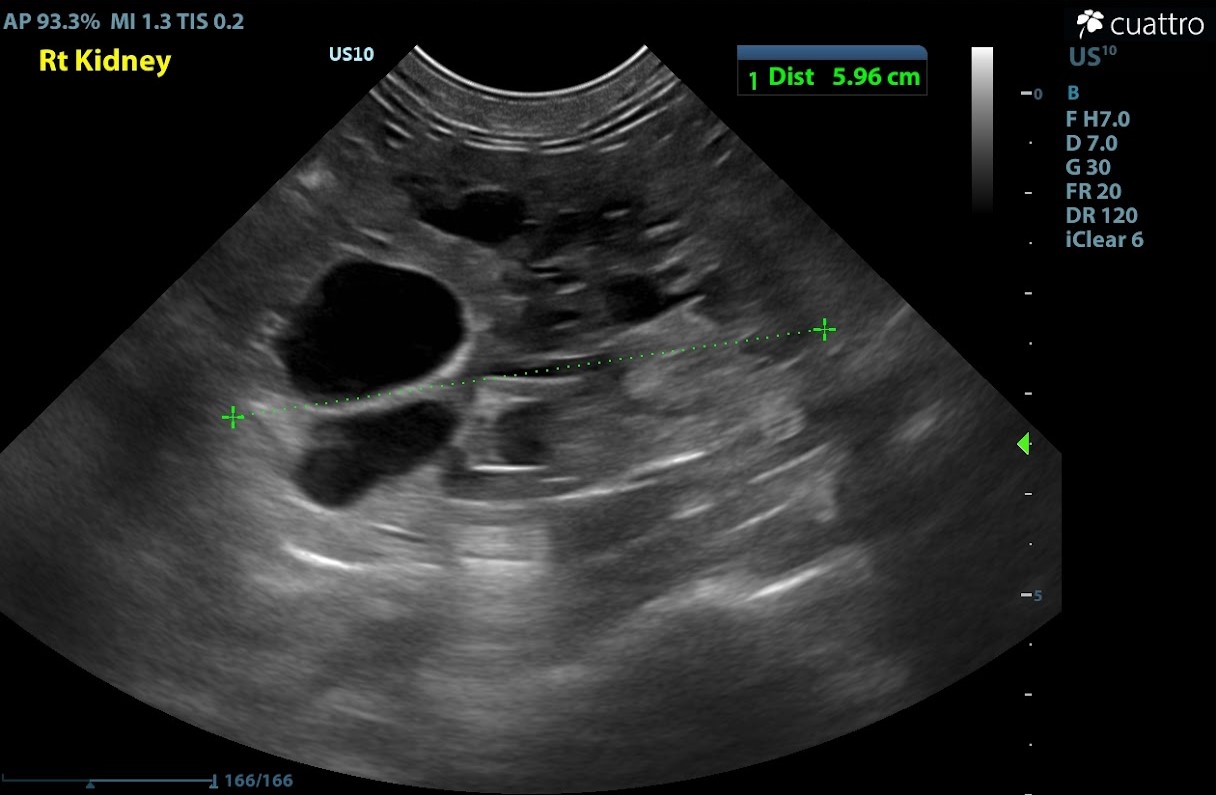
Sagittal orientation view of the right kidney displaying similarly increased kidney length, anechoic cortical cystic lesions and relatively normal overall shape.
Recommendations:
This patient appears to have a severe affliction of polycystic kidney disease bilaterally. Prognosis is considered poor to grave as this condition is progressive and generally ends with renal failure. As both kidneys are affected, there are limited options as far as surgical removal of diseased kidneys. In order to better determine a specific survival time expectancy for this patient, referral to a veterinary internal medicine specialist is recommended. Survival time is likely closer linked to individual patient condition, comfort and response to fluid therapy.
If further diagnostics are not pursued, consider palliative/symptomatic therapy to maintain quality of life.
Discussion:
Occurring most frequently in Persian cats, polycystic kidney disease is an inherited disorder in which small, closed, liquid-filled sacs develop in the tissue of the feline kidney. These sacs (cysts) tend to multiply in number and grow in size over time, eventually overwhelming normal kidney tissue and often leading to potentially fatal kidney failure. There is no explanation for the development of these cysts except for a genetic anomaly that is evident primarily in Persians and occasionally in a few other feline breeds, such as Himalayans and British Shorthairs.
In an affected cat, the cysts are present at birth and, though minuscule, may be diagnosed in kittens as young as six months of age. The number of these cysts varies from cat to cat, as does their size and the rate at which they grow within the kidney. The growths in a mature cat typically range in size from less than one millimeter to more than a centimeter in diameter. Clinical signs of polycystic kidney disease are most often recognized in cats that are seven years of age or so, although the disorder may be diagnosed in patients that are several years younger or older than that.
Patient outcome:
This patient was advised of recommendations by the primary care veterinarian. The last update regarding this patient was that the owners have elected to pursue palliative care to maintain the best quality of life for this pet. Understanding that this disease is individually progressive, there is not a specific timeline for survival.
Many thanks to our referring hospital, Kenhaven Animal Hospital, for entrusting us with their patient.