Ultra rare pancreatic disease in cats
Patient Information:
Age: 15 years, 9 months
Gender: Male Neutered
Breed: DLH
Species: Feline
History:
This patient has experienced vomiting, on and off, for the past 2 years. The patient was recently treated at an emergency facility for inappetence and frequent vomiting. Symptomatic therapy was initiated, and a recommendation for an abdominal ultrasound was made during the emergency visit.
Significant Ultrasonographic Findings:
Liver: Subjectively normal size, normal shape, and mildly hypoechoic echogenicity with coarse echotexture. No focal lesions are appreciated.
The gallbladder is moderately distended with anechoic and dense hyperechoic organizing bilious sludge adhering to the gallbladder wall. The gallbladder wall appears thickened with areas of severely hyperechoic comet-tails concerning for emphysematous cholecystitis. Significant common bile duct dilation is seen, with a maximum diameter of approximately 0.5 cm. The gallbladder is diffusely severely dilated to the level of the major duodenal papilla. A homogenous hyperechoic mass in the region of the major duodenal papilla measures approximately 0.4 cm in diameter.
Kidneys: Normal size (Lt/Rt = 3.2/3.3 cm) and shape with hyperechoic renal cortices, disproportionately enlarged, and decreased corticomedullary dimensions. No pyelectasia visualized.
Spleen: Enlarged size (1.2 cm at the hilus), shape, and echogenicity. No focal lesions appreciated.
Intestines: The stomach is empty and collapsed with normal rugal folds and layering. The pylorus is free of obstruction. Some small intestinal loops have prominent muscularis bowel layering, increased thickness, and subjectively normal motility. No loss of layering, obstruction, or masses seen. The colon has normal wall thickness and layering throughout.
Duodenum: 3.5 mm
Jejunum: 2.1-2.8 mm
Colon: 1.6 mm
Pancreas: Enlarged size, deranged shape, and heterogeneous hypoechoic echogenicity. The pancreatic tissue is diminished due to diffuse severe pancreatic duct dilation, with a max dilation of approximately 1.0 cm. There appears to be an irregular crystalline severely hyperechoic calculus within the lumen of the distal left pancreatic duct measuring 1.1 x 0.7 cm, along with smaller mineral opacities throughout the pancreas. Peripancreatic fat is severely hyperechoic.
Serosal surfaces: Mesentery throughout the abdomen is diffusely hyperechoic.
Abdominal Ultrasound Interpretation:
Liver: The findings are mild:
a) Chronic vs. Acute hepatitis or cholangiohepatitis (bacterial vs. sterile vs. toxin)
b) Steroid hepatopathy / Vacuolar hepatopathy / Glycogen storage disease
c) Infiltrative neoplasia (lymphosarcoma)
d) Hepatic lipidosis
Pancreas: Pancreatitis vs. pancreatic neoplasia (adenocarcinoma) vs. remotely abscess formation (pancreatic vs. free abdominal). Pancreatitis is likely in this case but pancreatic neoplasia cannot be ruled out.
Intestines: The findings are mild to moderate: Inflammatory bowel disease vs. infiltrative neoplasia (small-cell lymphosarcoma). Ultrasound cannot determine definitively whether the disease is benign or infiltrative.
[The combination of findings is suggestive of feline triaditis (inflammatory bowel disease, pancreatitis, and cholangiohepatitis).]
Common bile duct dilation: Severe findings: pancreatitis, cholangitis, hepatitis, duodenitis, neoplasia, cholelithiasis, fibrosis, or less likely mucus plugs, foreign bodies, or liver flukes.
Pancreaticolithiasis: Severe findings: chronic pancreatitis, pancreatic nodular hyperplasia, pancreatic pseudobladder, duplicate gallbladder, inflammatory bowel disease, and exocrine pancreatic insufficiency.
Splenomegaly: Mild findings: neoplasia (mast cell, lymphoma, carcinoma, metastatic disease) vs. fungal (Histoplasmosis) vs. reactive lymphoid hyperplasia vs. splenitis vs. congestion vs. extramedullary hematopoiesis (EMH).
Images:
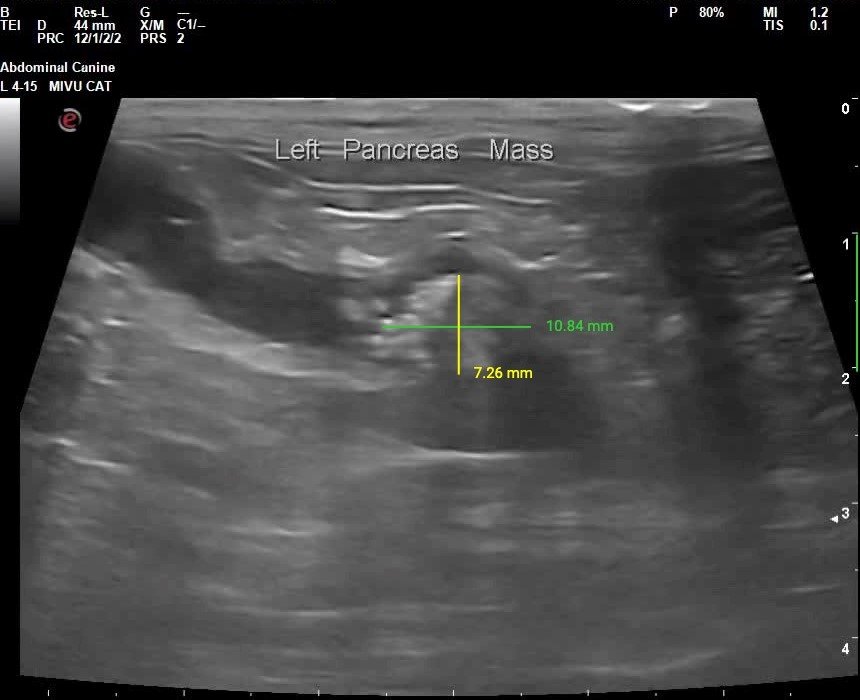
Image 1: Sagittal view of the distal left pancreas with mineralized structure(pancreaticolith) within the pancreatic duct.
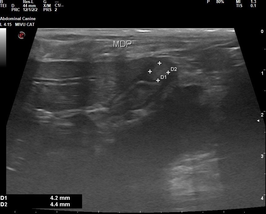
Image 2: Sagittal image of the major duodenal papilla which appears abnormally enlarged due to suspected mass effect.
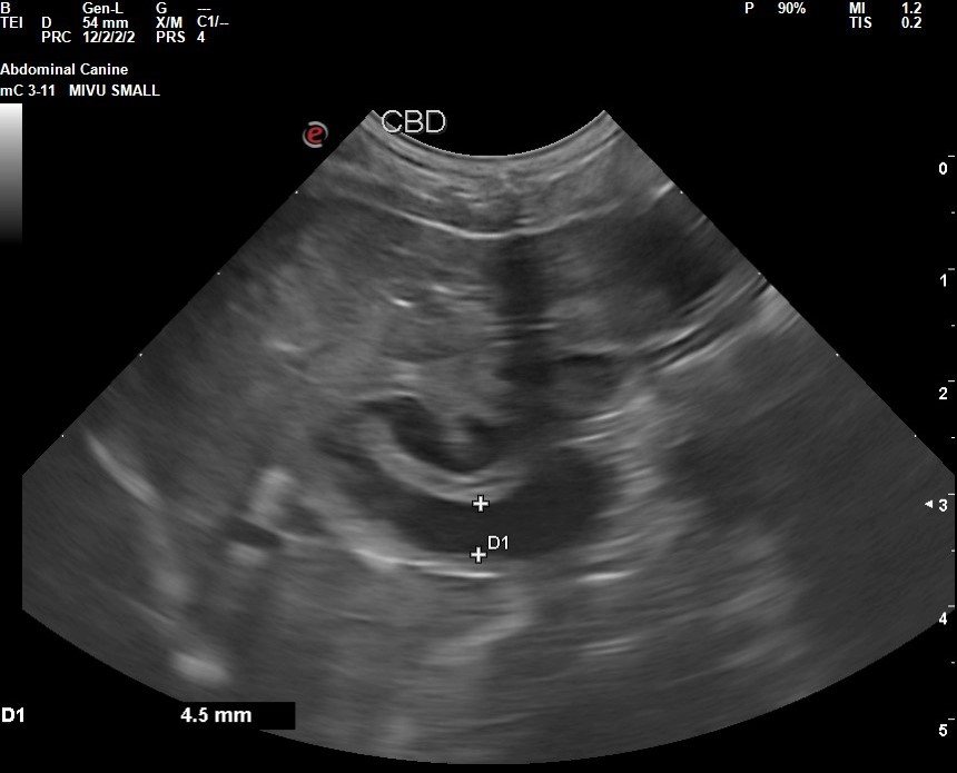
Image 3: Transverse view of the common bile duct which appears abnormally dilated. Dilation likely is secondary to blockage of flow at the level of the major duodenal papilla.
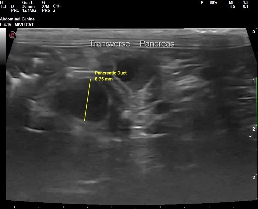
Image 4: Transverse image of the pancreatic body with severe duct dilation.
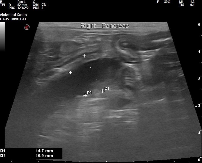
Image 5: Sagittal view of the right pancreas showing a severely dilated right pancreatic duct.
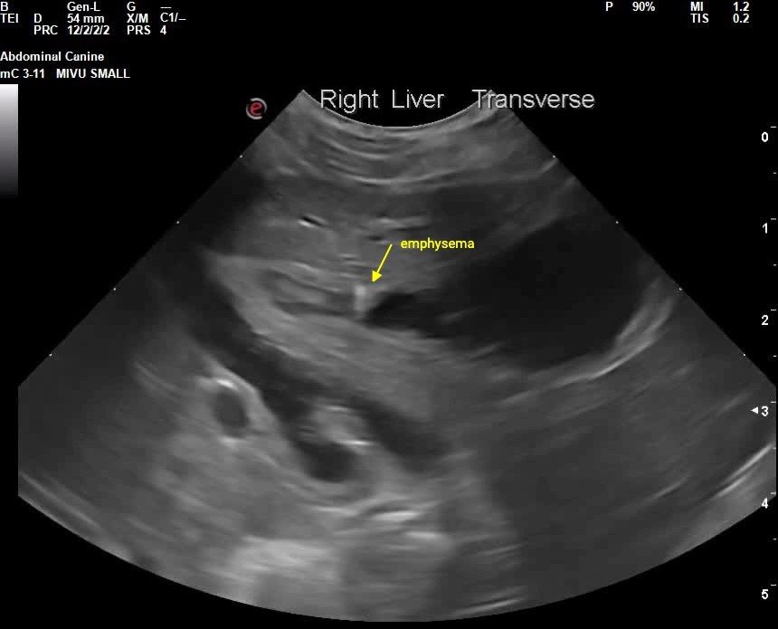
Image 6: Sagittal view of the gallbladder showing suspected gas infiltration of the gallbladder wall. Emphysematous cholecystitis is likely a sign of severity of infection in the gallbladder.
Recommendations:
- Referral to a veterinary surgeon for advanced imaging (e.g., CT scan) and discussion of therapeutic options is highly recommended.
- Cholecystocentesis with culture and sensitivity is recommended. Initiation of fluoroquinolone +/- metronidazole for suspected hepatitis/cholangitis is also recommended.
- It is NOT recommended to treat patients with evidence of common bile duct obstruction with Ursodiol.
- Appropriate pain medication, anti-emetics, and other supportive care therapy are advised.
- Thoracic radiographs (3-view, metastasis check) are recommended.
- If further diagnostics are not pursued, consider palliative or symptomatic therapy to maintain quality of life.
Discussion:
Pancreaticolithiasis is a well-documented condition in humans but has only been reported in 5 feline cases as of 2021. In humans, it is a sequela to chronic pancreatitis, occurring in 50–90% of cases. Cats share anatomical similarities, such as the pancreatic duct joining the common bile duct before entering the duodenum. Obstruction can result in ductal hypertension, leading to pain as the primary clinical sign.
Most feline pancreatoliths are composed of calcium carbonate, suggesting a similar pathomechanism to humans. Disease associations in felines include chronic pancreatitis, pancreatic nodular hyperplasia, inflammatory bowel disease, and exocrine pancreatic insufficiency.
Previously reported abnormalities in feline pancreatolithiasis include non-regenerative anemia, neutrophilia, and elevated liver enzymes, but normal ALP, GGT, and bilirubin levels. Hypercalcemia, while common in human cases, had not been reported in cats until recently.
Chronic pancreatitis is assumed to be the primary disease process, though acute hypercalcemia or ductal obstruction could exacerbate symptoms. Diagnosis often involves vague serum biochemistry results, with DGGR lipase offering high sensitivity for acute pancreatitis but low sensitivity for chronic cases.
Human treatments, such as extracorporeal shockwave lithotripsy and surgical removal, have shown success but are rarely available in veterinary medicine. Surgical removal in three cats has been described, with survival times ranging from 9 days to 2 years postoperatively.
Patient Outcome:
Recommendations were provided to the primary care veterinarian. Unfortunately, the owner could not be reached for follow-up, and the patient outcome remains unknown.
Sonographer:
Mallory Repellin, DVM
A very special ‘thank you’ to Dr. Julie Augustine from Montgomery Animal Hospital for sharing patient details and allowing for an in-depth discussion of their patient.