Suspected Carcinomatosis in a Feline
Patient Information:
Age: 11 years
Gender: Spayed Female
Breed: Himalayan
Species: Feline
History:
Patient presented with a history of diarrhea and significant weight loss. Laboratory work revealed severe neutrophilic leukocytosis, non-regenerative anemia, and hypoalbuminemia.
Abdominal Ultrasonographic Findings:
The aboral ileum and oral ascending colon are severely thickened (up to 7.2mm) and hypoechoic, with complete loss of normal layering distinction. This creates a mass effect measuring 3.5x2.5cm at the ileocecocolic junction.
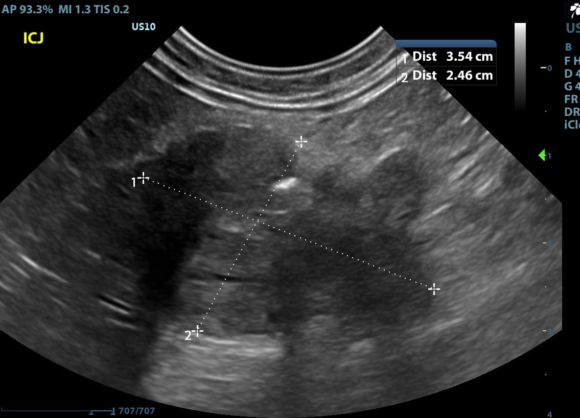
Prominent (1.0cm diameter) rounded hypoechoic mesenteric lymph nodes are noted in the area of the ICCJ mass.
A scant amount of anechoic peritoneal fluid is present.
The mesentery is moderately-severely hyperechoic surrounding the ICCJ mass.
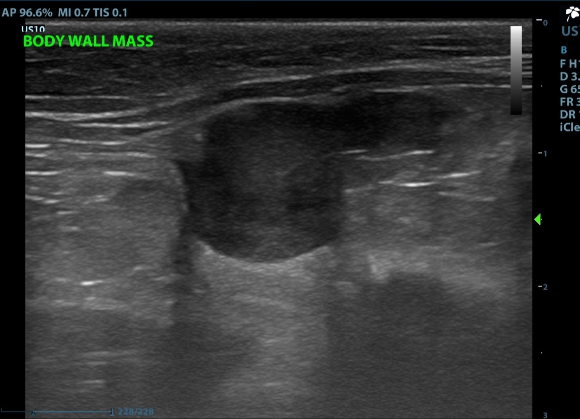
A homogenous rounded hypoechoic mass (1.2x1.1cm) is present that appears to originate from the peritoneal wall and extends into the peritoneal cavity.
Ultrasound Interpretation:
-
Ileocecocolic Mass: The findings are severe.
DDx: Adenocarcinoma, infiltrative neoplasia (lymphosarcoma, mast cell tumor), feline gastrointestinal eosinophilic sclerosing fibroplasia (FGESF), leiomyosarcoma, leiomyoma. -
Lymph Nodes: The findings are moderate.
DDx: Reactive, infection, infiltrative neoplasia (lymphoma, mast cell, other), metastatic neoplasia. -
Body Wall Mass: The findings are moderate.
DDx: Metastasis (carcinomatosis), granuloma, other. -
Ascites: This finding is very mild.
DDx: Transudate, hemorrhagic, exudate.
Diagnostic Recommendations:
While disseminated disease was suspected, ultrasound-guided fine needle aspiration was recommended to arrive at a more definitive diagnosis.
Cytology Results:
Diagnosis
ICCJ Mass: Suspect carcinoma.
Body Wall Mass: Suspect carcinoma.
Comments
For both locations, a carcinoma is suspected with 80% confidence, suggestive of a metastatic process. However, due to a lack of convincing criteria of malignancy, the possibility of two benign epithelial proliferations cannot be completely ruled out. Consider biopsy/histopathologic evaluation.
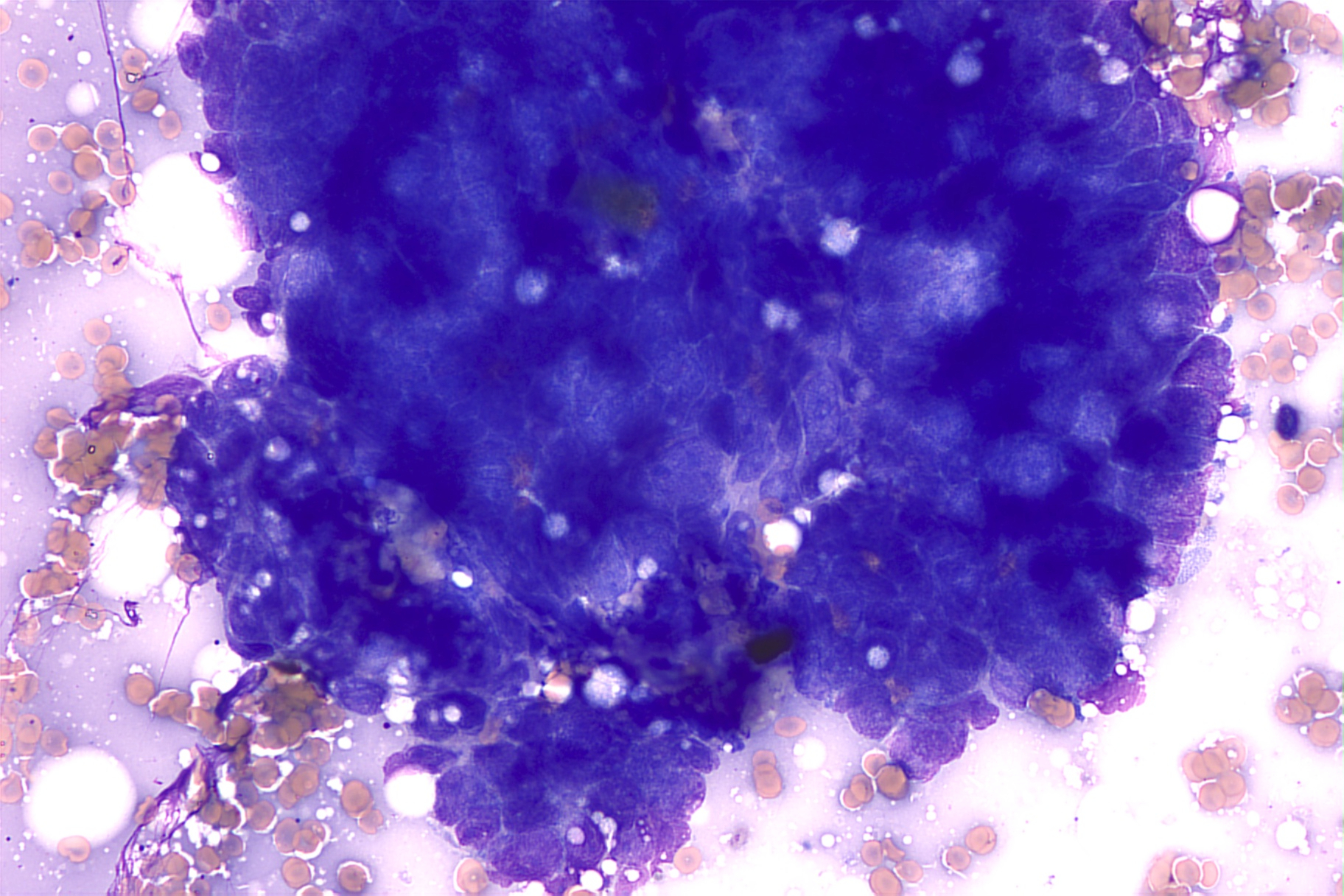
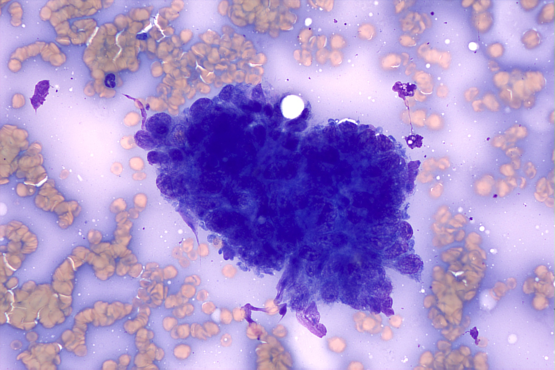
Microscopic Description
The slides from the two locations appear very similar, are moderately cellular, and consist of a small amount of cellular debris, few to many red blood cells, and a nucleated cell population predominated by mature leukocytes in proportions consistent with the peripheral blood present. Fewer round to polygonal cells are found in cohesive clusters. These cells have a small amount of very basophilic cytoplasm surrounding a central, round nucleus with a coarsely-stippled chromatin pattern and occasional visible nucleoli. Anisokaryosis is mild to moderate.
Casey J. LeBlanc, DVM, PhD, DACVP (Clinical Pathology)
Recommendations:
Carcinomatosis was the most likely diagnosis given the full clinical picture. Oncology consult was recommended for an optimal diagnostic and treatment plan versus palliative care or humane euthanasia. Prognosis was very guarded given the current information.
Patient Outcome:
Palliative care was initially elected, but due to continued declining condition, the owner elected humane euthanasia a short time later.
Discussion:
Carcinomatosis is the widespread dissemination of tumor cells in a body cavity (often peritoneal). This is most often associated with epithelial cell tumors but can also occur with sarcoma or lymphoma. The most common primary tumor sites are the pancreas, gastrointestinal tract, and hepatobiliary system. It is important to note that a primary tumor is not always identified. Ascites of varying echogenicity is typically present, along with hypoechoic nodules within the mesentery. In some cases, as in this one, nodules can occur on the body wall. Lymphadenopathy is also common.
Diagnosis can be challenging; biopsy and histopathology may be needed for a definitive diagnosis. However, as in this case, less invasive ultrasound-guided fine needle aspirates can sometimes yield results and should be discussed when the suspicion of carcinomatosis is present.
Unfortunately, limited treatment options exist for carcinomatosis, and treatment is often palliative. This may involve periodic abdominocentesis to relieve discomfort, anti-nausea medication, and pain control, as this condition is often described as painful in humans. Intracavitary carboplatin has been attempted, but no studies have demonstrated efficacy in cats, and it remains only a palliative option at this time.
References:
Monteiro CB, O’Brien RT. A retrospective study on the sonographic findings of abdominal carcinomatosis in 14 cats. Vet Radiol Ultrasound 2004; 45: 559–564.
Weston PJ, Baines SJ, Finotello R, et al. Clinical, CT, and ultrasonographic features of canine and feline pleural and peritoneal carcinomatosis and sarcomatosis. Vet Radiol Ultrasound 2021; 62: 331–341.
Sonographer:
Kara Woody, DVM
Special thanks to Burke Veterinary Clinic and Eastern VetPath for collaboration on this sad but interesting case!