Lameness in a Dog
Patient Information:
Age: 4 years old
Species: Canine
Breed: Labrador Retriever
Gender: Spayed Female
History:
The patient presented for evaluation of a 6-week history of left thoracic limb lameness. No fall or trauma was reported. The lameness worsened with exercise. On physical examination, the patient showed a grade 1/5 LF lameness characterized by short striding at a walk and trot with some scuffing, and she was off-weight at a stand. Discomfort was noted on flexion and extension of the shoulder, and tension was detected on a biceps tendon stretch.
Radiographs LF:
An increased soft tissue opacity was visualized within the bicipital groove.
Ultrasound Findings:
Ultrasound evaluation of the LF shoulder was performed with the right side for comparison. The distal portion of the infraspinatus tendon (IST) was moderately enlarged, measuring 6.9 mm in thickness and 0.74 cm 2 in cross-section, compared to the right (2.8 mm thick). The affected portion of the tendon was composed of disrupted hypoechoic irregular fibers and lacked the normal longitudinal hyperechoic fiber pattern. A few hyperechoic pinpoint areas of dystrophic mineralization were observed in the affected portion of the tendon. The hyperechoic bone surface of the humeral head at the attachment of the IST appeared slightly irregular. The infraspinatus muscle had a normal sonographic appearance.
A large amount of anechoic fluid was present in the infraspinatus bursa, measuring 0.54 cm 2. The origin/proximal portion of the left biceps tendon had a mildly irregular contour and contained a few small hypoechoic areas of short, irregular fibers. The wall of the biceps tendon sheath was slightly thickened (1.5 mm) and hyperechoic. A mild to moderate amount of anechoic fluid was seen in the distal outpouching of the biceps tendon sheath, measuring 0.20 cm 2.
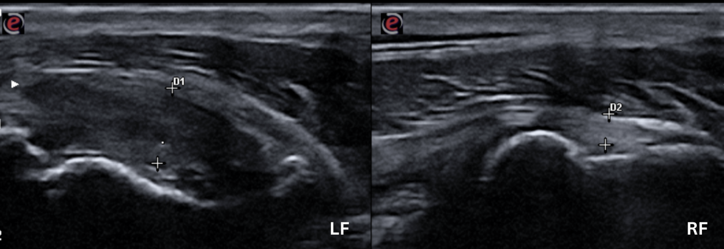
Image 1. Longitudinal view of the of the LF and RF infraspinatus tendons. The LF IST is enlarged, irregular and hypoechoic, lacking normal hyperechoic fibrillar pattern compared to the RF IST.
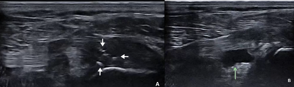
Image 2. A. Longitudinal view of the LF IST showing the hyperechoic shadowing foci of dystrophic mineralization present within affected tendon (arrows). B. Longitudinal sonogram showing a large amount of anechoic effusion within the infraspinatus bursa (green arrow).
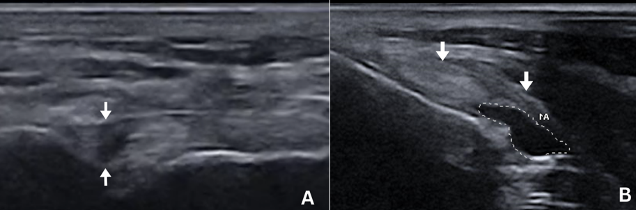
Image 3. A. Longitudinal sonogram showing the hypoechoic irregular fibrillar pattern at the origin of the biceps tendon (arrows). B Thickening (arrows) and anechoic effusion (dotted line) within the biceps tendon sheath.
Diagnosis:
Severe acute LF infraspinatus tendinopathy/partial rupture, moderate to severe infraspinatus bursitis, mild biceps tendinopathy, and mild to moderate synovitis of the bicipital tendon sheath.
Treatments:
An intra-articular injection of platelet-rich plasma (PRP) and a total of 3 extracorporeal shock wave therapy sessions were performed on the left shoulder. Oral medications included codeine, methocarbamol, and carprofen, as well as glucosamine, chondroitin, and omega-3 fatty acid supplementation. A rehabilitation program was instituted, including manual, laser, and therapeutic ultrasound treatments. Exercise restriction was implemented for the duration of the dog’s recovery. Repeat ultrasound evaluation was recommended in 8 to 12 weeks to assess the progression of the injuries.
Update:
The patient is progressing well with full LF weight bearing at rest and no evidence of lameness at the walk. Physiotherapy and exercise restriction are still being recommended at this point of the healing process.
Injuries to the tendon of insertion of the infraspinatus muscle have been poorly described in veterinary medicine. According to the current literature, the incidence of infraspinatus tendinopathies in Labrador Retrievers is higher than in other breeds. 1 Most reported cases were treated with conservative therapy (NSAIDs and restricted exercise) and became asymptomatic several months following the initial injury. 1 Infraspinatus contracture is the primary myopathy that can potentially develop secondary to acute trauma to the infraspinatus muscle and/or tendon. 2,3
References:
- Mikola K et al. Isolated avulsion of the tendon of insertion of the infraspinatus and supraspinatus muscles in five juvenile Labrador Retrievers. Vet Comp Orthop Traumatol. 2018;31(4):285-290.
- Devor M et al. Fibrotic contracture of the canine infraspinatus muscle. Vet Compar Orthop Traumatol. 2006;19(4):117-21.
- Talor J et al. Acquired muscle contractures in the dog and cat. A review of the literature and case report. Vet Compar Orthop Traumatol. 2007;20(2):79-85.
Special thanks to the staff at Skylos Sports Medicine and Dr. Hummel for their help with this case.