Intestinal Mass in Dog
History: A 12-year-old spayed female Pomeranian mix presented for evaluation of chronic cough and polyuria/polydipsia. Blood work revealed an increase in alkaline phosphatase (561 U/L). Urinalysis showed a specific gravity of 1.019, 1+ protein, and 1+ blood. No gastrointestinal signs were reported.
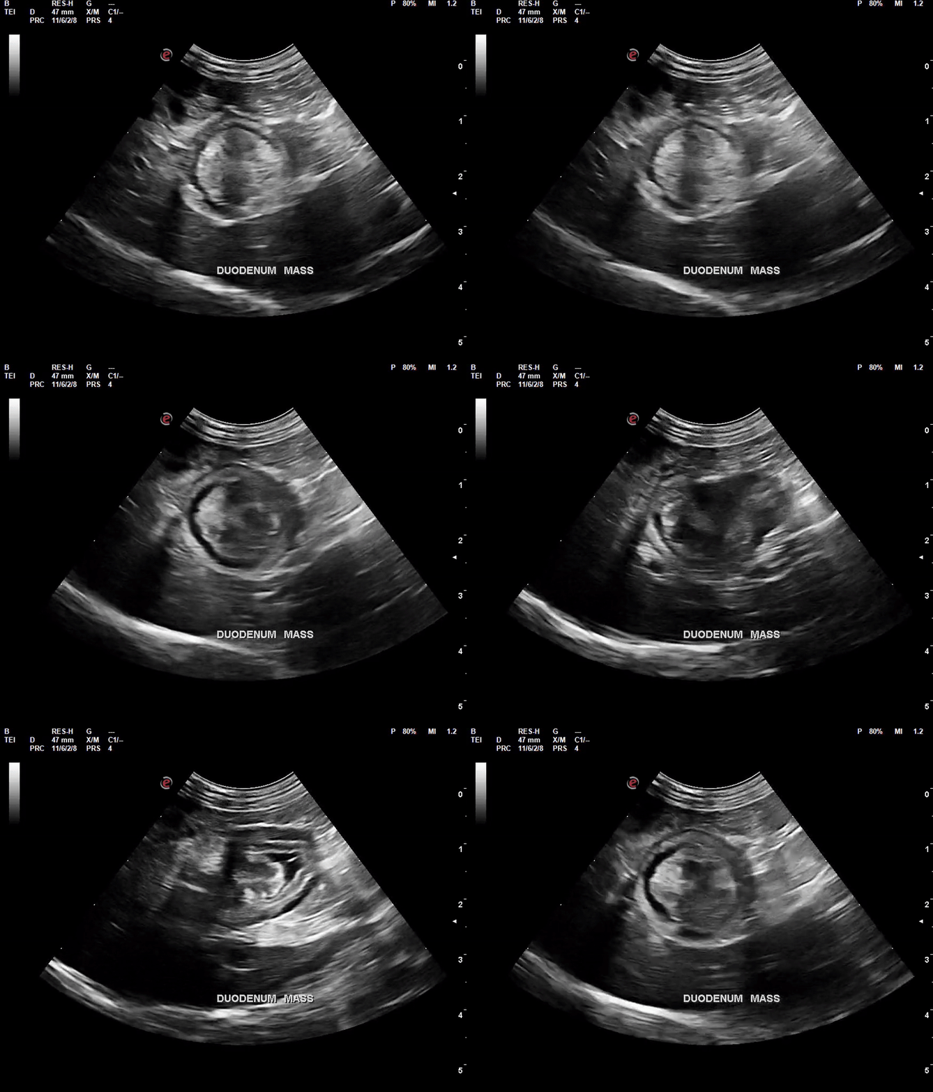
Ultrasound findings (initial): An ill-defined focal homogeneous hyperechoic non-obstructive mass was observed originating from the wall of the proximal duodenum, protruding toward the lumen. The lesion measured 1.0x1.5cm, with a decreased definition of normal wall layering in the affected duodenal segment. No abnormalities were noted in the rest of the small intestine or colon. Additional findings consistent with hyperadrenocorticism were noted, including hepatic lipidosis, gallbladder sludge, and unilateral adrenal enlargement. Urinary system changes included renal diverticular mineralization and cystoliths.

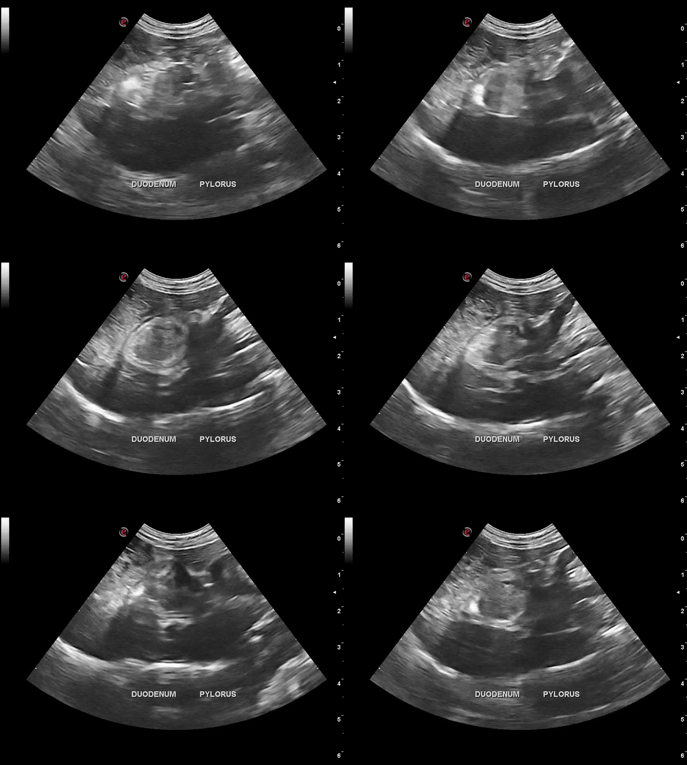
Video # 1. Ultrasound of the proximal duodenum showing the focal mass protruding into the duodenal lumen.
Recommendations: Endoscopic evaluation and biopsies of the duodenal lesion were discussed with the owner, but declined at this time. Repeat ultrasound was recommended in 6 to 8 weeks or sooner if gastrointestinal signs develop. Further testing for hyperadrenocorticism was advised, along with uroliths removal and management of secondary urinary tract infection. The owner opted for medical management of suspected bronchitis with prednisolone but declined further diagnostics or treatment at this stage.
Ultrasound findings (6 weeks later): the duodenal lesion had increased in size, now measuring 1.7x1.7cm and extended into the pylorus/pyloric antrum, where it measured 1.0x2.1cm. All other ultrasound findings remained stable, except for progression to bilateral adrenal enlargement.

Video #2. Ultrasound now showing the lesion extending into the pylorus/pyloric antrum.
Recommendations: due to the progression of the intestinal lesion, specialty referral for endoscopy and biopsies was again recommended. Instead, exploratory surgery was performed at another general practice to obtain incisional biopsies.
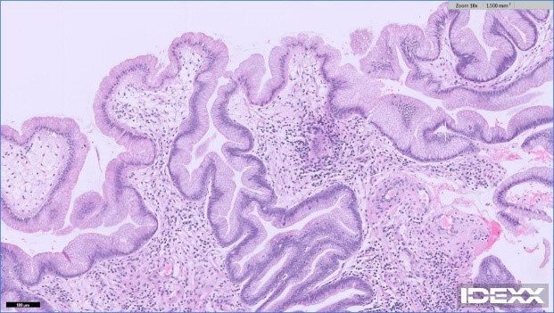
Histopathology results: findings were consistent with a benign hyperplastic intestinal polyp.

Images# 1: proliferation of epithelial cells into villous folds, with associated mild inflammation (lymphocytes, plasma cells, neutrophils). No evidence of malignancy noted.
Outcome: A follow-up ultrasound performed eight weeks post-surgery showed a slight reduction in the lesion size, likely attributable to prior tissue sampling. The patient remains asymptomatic.
Hyperplastic duodenal polyps are rare in dogs, with only one reported case causing hematemesis and melena1. They are also infrequently found in other locations of the canine gastrointestinal tract, including the stomach, usually as incidental findings, though occasionally linked to anemia or obstruction2-3. In the colon and rectum, only two cases were noted among 80 dogs with colorectal epithelial tumors4. While their significance remains unclear, hyperplastic polyps in humans may become obstructive or undergo neoplastic transformation5. Surgical removal is preferred for symptomatic or obstructive cases, with endoscopic resection an option for smaller lesions. Histopathology is essential to confirm benignity. Asymptomatic or non-resectable cases require close monitoring for progression.
- Patouchas O et al. Large duodenal hyperplastic polyp causing haematemesis and melena in a dog. Vet Rec Case Rep. 2024;12:e1005.
- Kim K et al. Gastric hyperplastic polyp causing upper gastrointestinal hemorrhage and severe anemia in a dog. Vet Sci. 2022(12):680.
- Kuan S et al. Ultrasonographic and surgical findings of a gastric hyperplastic polyp resulting in pyloric obstruction in a 11-week-old French bulldog. Aust Vet J, 2009;87(6): 253-55.
- Wolf JC et al. Immunohistochemical detection of p53 tumor suppressor gene in canine epithelial colorectal tumors. Vet Pathol. 1997;34(5(: 394-404.
- Jass JR. Hyperplastic polyps and colorectal cancer: is there a link? Clin Gastroenterol Hepatol. 2004;2(1):1-8.
Special thanks to Dr. Olech and the staff at Piedmont Pets Veterinary Care for their help with this case.

