Suspected Carcinomatosis in a Geriatric Canine
Patient Information:
Age: 11 years
Gender: Spayed Female
Breed: Mixed
Breed Species: Canine
History:
Acting weak and lethargic at home.
Elevated ALP and non-regenerative anemia.
Possible space occupying mass in cranial abdomen on radiographs.
Abdominal Ultrasonographic Findings:
Liver: There is a contour deforming rounded mildly hypoechoic homogenous mass present in the medial left liver measuring 4.4x4.3cm-sagittal and 5.9x4.3cm-transverse. The tissue surrounding this mass is moderately-severely hyperechoic. The rest of the liver is normal size, shape and subjectively hypoechoic echogenicity.
Serosal surfaces: The mesentery is diffusely hyperechoic and nodular particularly in the cranial abdomen where ill-defined hypoechoic small nodules are noted in the area of the pancreas. There is a small amount of echogenic peritoneal fluid noted in the caudal abdomen.
Ultrasound Interpretation:
Liver Mass - the findings are moderate - DDx: primary hepatic neoplasia vs. metastatic neoplasia vs. infiltrative neoplasia (lymphosarcoma) vs. benign nodules of regeneration.
Mesentery - the findings are moderate - DDx: metastasis (e.g. carcinomatosis, lymphomatosis vs sarcomatosis) vs peritonitis - inflammation vs. paraneoplastic reaction vs. infectious vs. fibrosis vs. other.
Ascites - this finding is moderate - DDx: transudate vs. hemorrhagic vs. exudate
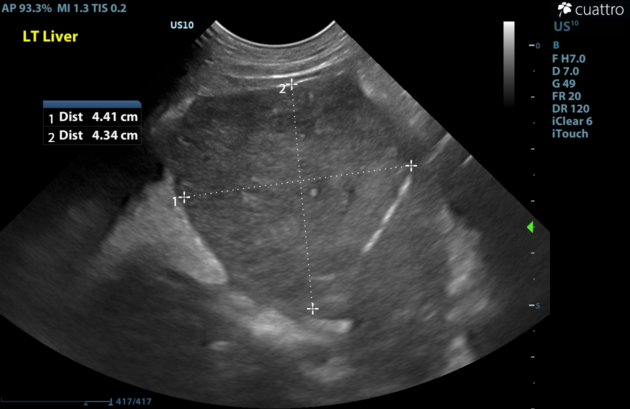
Image 1: Rounded mildly hypoechoic mass on the left medial liver.
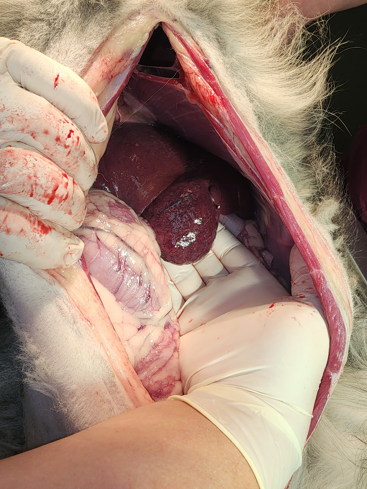
Image 2: Rounded mass on the left medial liver found on necropsy.
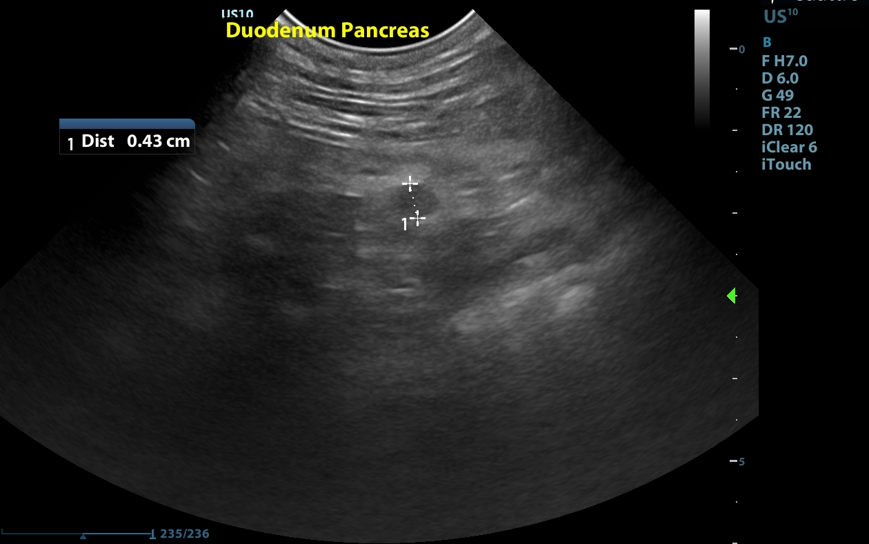
Image 3: A small hypoechoic nodule noted near the pancreas.
Diagnostic Recommendations:
Peritoneal fluid cytology and analysis as well as fine needle aspirate samples of the liver mass could be considered to further define. Carcinomatosis was most suspected with the primary tumor in the liver
Patient Outcome:
The owner elected humane euthanasia due to poor prognosis. Necropsy was performed.
Necropsy Results:
A rounded liver mass was found on the medial left liver lobe. TNTC small pale nodules were found throughout the mesentery and pleural cavity. There was also a small amount of serosanguinous peritoneal fluid present.
Discussion:
Histopathology was not performed in this patient since necropsy was mainly performed for clinical interest. However, the clinical findings of a liver mass as well as numerous mesenteric nodules and bicavitary effusion makes carcinomatosis the presumptive diagnosis. Carcinomatosis is the widespread dissemination of tumor cells in a body cavity (often peritoneal). This is most often from an epithelial cell tumor, but can also occur with sarcoma or lymphoma. The most common primary tumor sites are pancreas, gastrointestinal tract and hepatobiliary system, as was the case with this patient. It is also important to note that a primary tumor is not always identified. Peritoneal effusion of varying echogenicity is typically present as well as hypoechoic nodules within the mesentery. In some cases, nodules can occur on the body wall. Lymphadenopathy is also common.
Diagnosis can be challenging; biopsy and histopathology may be needed for definitive diagnosis. However, sometimes less invasive methods such as ultrasound guided fine needle aspirate of primary tumor and/or peritoneal fluid cytology can yield results and should be discussed when the suspicion of carcinomatosis is present.
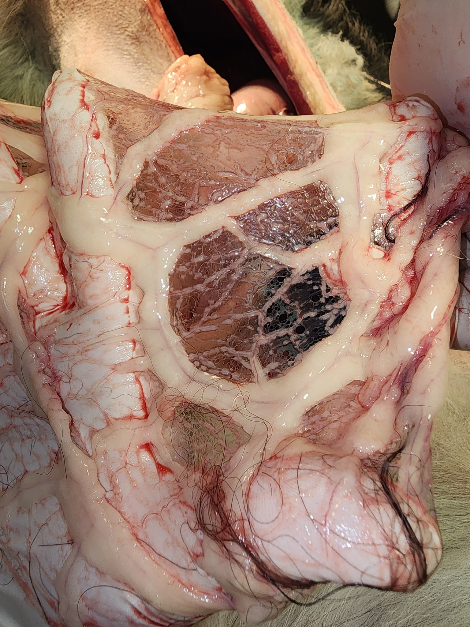
Image 4: TNTC small pale nodules seen throughout the mesentery found on necropsy.
Image 5: Serosanguinous peritoneal fluid found on necropsy
Unfortunately, limited treatment options exist for carcinomatosis and the treatment is often palliative involving periodic abdominocentesis to relieve discomfort, anti-nausea medication and pain control as this condition is often described as painful in humans. Intracavitary mitoxantrone and/or carboplatin/cisplatin has been described and while generally well tolerated, results are mixed and reliable efficacy has not been established. A recent study showed some promise with oral Palladia, but larger studies are needed.
References:
Clinical, CT, and ultrasonographic features of canine and feline pleural and peritoneal carcinomatosis and sarcomatosis. Vet Radiol Ultrasound 2021; 62: 331–341.
Evaluation of intracavitary mitoxantrone and carboplatin for treatment of carcinomatosis, sarcomatosis and mesothelioma, with or without malignant effusions: A retrospective analysis of 12 cases (1997-2002). Vet Comp Oncol. December 2005;3(4):171-81.
Retrospective evaluation of toceranib phosphate (Palladia) in the treatment of canine carcinomatosis and mesothelioma Vet Comp Oncol. June 2024;22(2):245 - 254
Sonographer: Kara Woody, DVM
Special thanks to Stafford Animal Hospital for collaboration on this interesting, but sad case.